IMAGE

FIGURE 5

ID
ZDB-IMAGE-210307-48
Source
Figures for Hu et al., 2021
Image
Figure Caption

FIGURE 5

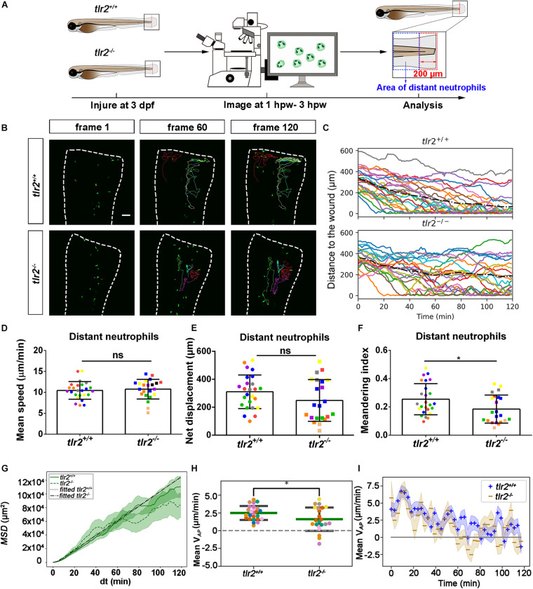
Quantification of distant neutrophils behavior in wounded tlr2 mutant and sibling control larvae. (A) Experimental scheme. Tlr2+/+ and tlr2– /– larvae were wounded at 3 dpf. The red dashed line shows the site of wounding. Neutrophils of wounded zebrafish larvae were tracked for 2 h and images were taken every 1 min by using CLSM. For cell tracking analysis, cells localized outside an area of 200 μm from the wounding edge toward the body trunk were counted as distant cells. Blue dashed box shows the area where distant neutrophils were tracked. (B) Representative images of distant neutrophil tracks in the wounded tail fin of 3 dpf tlr2+/+ or tlr2– /– larvae at frame 1, frame 60 and frame 120. Time interval between two successive frames is 1 min. Each color track represents an individual neutrophil. Cell tracking movies are shown in Supplementary Movies S9, S10). Scale bar: 50 μm. (C) Distance to the wound. Black dash line represents average distance to the wound. Each color line represents one cell. (D–I) Quantification of distant neutrophil tracks. In (D–F,H), each color indicates a different larva. There was no significant difference between the groups in terms of mean speed (D), net displacement (E), and MSD (green) and fitted MSD (black) (G). However, meandering index (F) and mean VAP(H) of neutrophils at the wound in tlr2+/+ is greater than in tlr2– /– larvae. The fitted MSD (G, black) was fitted for dt < 80 min. The shaded regions in MSD (G) and mean VAP over time (I) indicate standard error of the mean. Statistical analyses were done with 7 and 8 fish, respectively, for each group. An unpaired, two-tailed t-test was used to assess significance (ns, non-significance, *P < 0.05) and data are shown as mean ± SD. Sample size (n): 25, 22 (D–F,H).

Acknowledgments
This image is the copyrighted work of the attributed author or publisher, and ZFIN has permission only to display this image to its users. Additional permissions should be obtained from the applicable author or publisher of the image. Full text @ Front Cell Dev Biol