
|
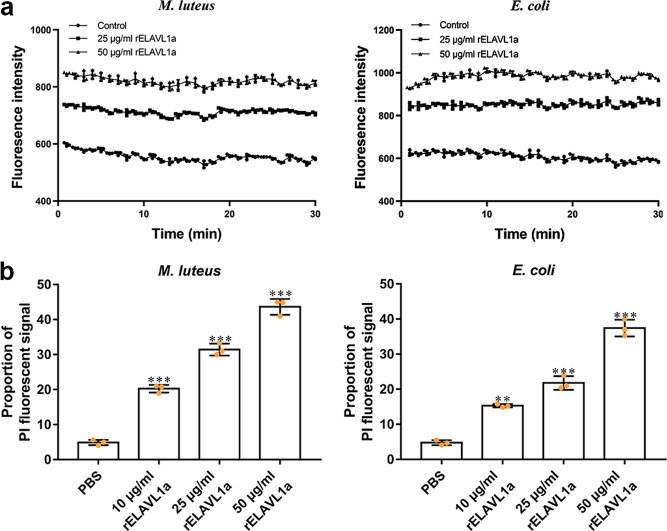
Fig. 5 a The rELAVL1a caused depolarization of the bacterial plasma membrane. The changes in fluorescence intensity were recorded with a Tecan GENios plus spectrofluorometer at an excitation wavelength of 622 nm and an emission wavelength of 670 nm. Control, HEPES buffer containing 20 mM glucose. b The effects of rELAVL1a on the membrane integrity of M. luteus and E. coli, cells analyzed by flow cytometry. All data were expressed as mean values ± SD. (n = 3). The data are from three independent experiments performed in triplicate. The bars represent the mean ± S.D. The significance of the difference was determined by one-way ANOVA. **p < 0.01, ***p < 0.001.