
Image
|
Figure Caption
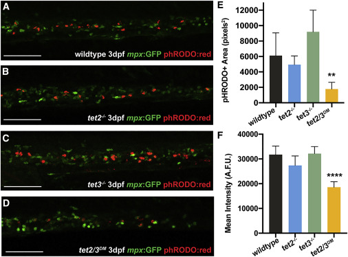
Fig. 3 Figure 3. Phagocytosis Defect Is Unique to tet2/3DM Embryonic Neutrophils (A–D) Representative images of the CHT region in pHrodo-red-injected 3-dpf wild-type, tet2−/−/, tet3−/−, and tet2/3DM mpx:GFP embryos. Scale bars represent 200 μm. (E) Quantification of phagocytosed beads by fluorescent area within GFP+ neutrophils in pixels squared. (F) Quantification of lysosome acidification by mean fluorescence intensity in arbitrary fluorescence units (A.F.U.). n = 5–7 embryos/condition, ∗∗p < 0.01, ∗∗∗∗p < 0.0001 by 2-way ANOVA with Tukey post hoc test of each condition compared to wild type.
Acknowledgments
This image is the copyrighted work of the attributed author or publisher, and
ZFIN has permission only to display this image to its users.
Additional permissions should be obtained from the applicable author or publisher of the image.
Full text @ Cell Rep.