
Image
|
Figure Caption
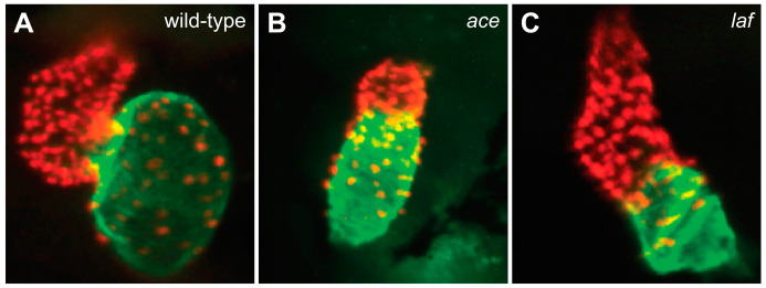
Figure 3 FGF and BMP signaling promote ventricular and atrial cardiomyocyte formation, respectively. Compared to the chambers of the wild-type heart at 48 hpf (A), the ace (fgf8a) mutant heart (B) exhibits a substantially reduced ventricle, and the laf (acvr1l) mutant heart (C) exhibits a substantially reduced atrium. Red fluorescence labels cardiomyocyte nuclei, and green fluorescence indicates localization of Amhc. Images adapted from [54,59].
Acknowledgments
This image is the copyrighted work of the attributed author or publisher, and
ZFIN has permission only to display this image to its users.
Additional permissions should be obtained from the applicable author or publisher of the image.
Full text @ J Cardiovasc Dev Dis