
|
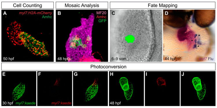
Figure 1 Useful tools for analysis of cardiac chamber development in zebrafish. (A) Transgenes such as Tg(myl7:H2A-mCherry) label myocardial nuclei (red) and facilitate counting of the cardiomyocytes in each chamber; the atrial myosin heavy chain Amhc (green) distinguishes the atrium from the ventricle. Lateral view of wild-type heart at 50 h post-fertilization (hpf) is adapted from [27]. (B) Mosaic analysis enables assessment of the cell-autonomy of gene function. In this example, adapted from [27], mosaic distribution of the transgene Tg(hsp70:dnfgfr1-eGFP) (green) inhibits FGF signaling in specific ventricular cells, several of which exhibit ectopic Amhc, indicating a cell-autonomous requirement for FGF signaling to repress amhc expression in the ventricle (lateral view). Magenta fluorescence labels sarcomeric myosin heavy chain using the monoclonal antibody MF20, and red fluorescence indicates localization of Amhc, using the monoclonal antibody S46. (C,D) Fate mapping follows progenitor cells from their origins to their destinations. In this example, adapted from [28], photoactivation of a caged fluorescein-dextran lineage tracer marks a small group of cells in the anterior lateral plate mesoderm (ALPM) at the 6–9 somite (som) stage (C, dorsal view). Later, labeled progeny of these cells (arrowheads) are found in the atrium; uncaged fluorescein (blue) is detectable within the myl7-expressing myocardium (magenta) of the heart (D, frontal view). (E–J) Photoconvertible proteins facilitate tracking of cells over time. In this example, adapted from [29], cardiomyocytes express the transgene Tg(myl7:kaede), and regionally restricted photoconversion of Kaede at 30 hpf converts its green fluorescence into red fluorescence near the arterial pole of the heart tube (E–G). Later, visualization of retained red fluorescence demonstrates that the labeled cells contribute to the ventricle (H–J).