
|
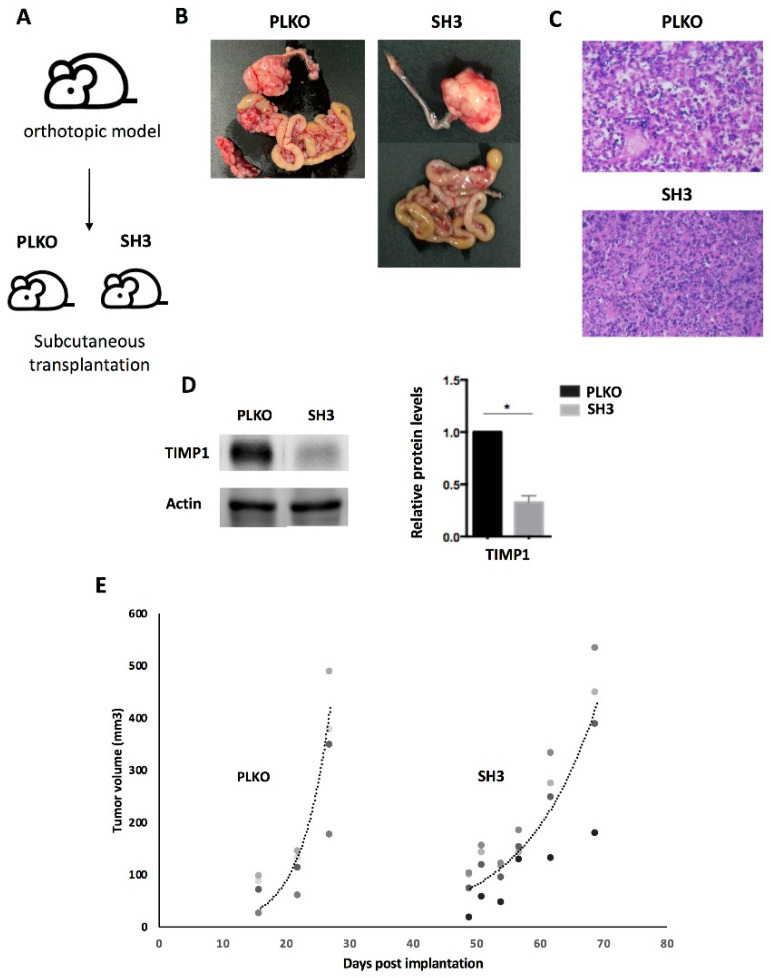
Figure 6 Effect of TIMP1 knock-down in SKOV3 proliferation in a mice model. (A) Representative scheme of the assay. The TIMP1 silenced cells (SH3) and the control cell line (PLKO) were injected in the ovary of SCID mice and the generated tumors amplified subcutaneously in the flank of three female SCID mice per group. (B) Macroscopic image of tumor generated orthotopically. (C) Representative images of hematoxylin-eosin staining of tumors generated in the subcutaneous model (objective 10x). (D) Western blot results for TIMP1 protein levels in tumors generated by SKOV3_PLKO and SH3 cell lines. Actin was used as normalizer. (E) Dynamic of tumor growth after subcutaneous transplantation, which was slower in the TIMP1 silenced cell line. nreplica = 3 for the two experimental groups. * p < 0.05.