
|
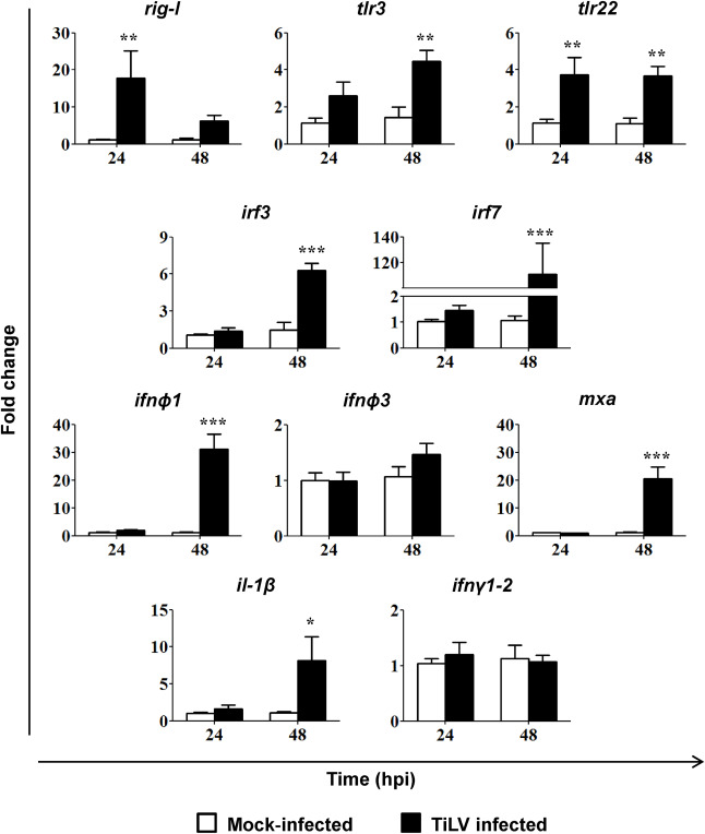
Fig. 3 Fig. 3. TiLV-induced changes in the expression of genes involved in antiviral response. Gene expression of the pathogen recognition receptors (rig-I (ddx58), tlr3, tlr22), transcription factors (irf3, irf7), type I interferon (infϕ1, infϕ3), antiviral protein (mxa) and pro-inflammatory cytokines (il-1β, ifnγ1-2) in 5 pooled larvae of zebrafish mock-infected or TiLV-infected by injection into the duct of Cuvier at 2.5 dpf. The gene expression is normalized against the housekeeping gene rps11. Changes in gene expression of TiLV-infected larvae (black bars) are shown as x-fold increase compared to mock-infected control larvae (white bars) at each time point. The symbol (*) indicates significant differences between the mock-infected and TiLV infected larvae at each time point (*p ≤ 0.05; **p ≤ 0.01; ***p ≤ 0.001) as revealed by two-way ANOVA followed by a Bonferroni test. Each bar represents the mean + SD of n = 7–8 samples derived from two independent experiments.