
|
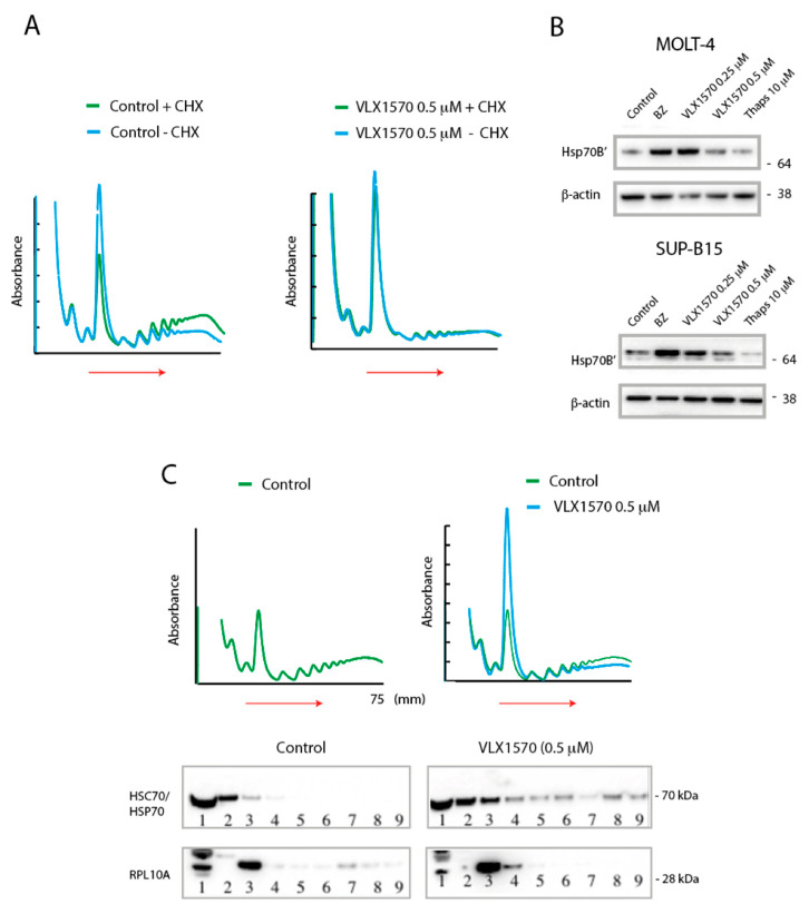
Figure 5 Evidence for impaired translational elongation. (A) SUP-B15 cells were exposed to VLX1570 or vehicle and extracts fractionated by sucrose gradient centrifugation. Extracts were prepared in the presence or absence of cycloheximide (CHX) as indicated. (B) MOLT-4 or SUP-B15 cells were exposed to the indicated compounds for 6 h, and cell extracts were processed for immunoblotting. Bortezomib (BZ) was used at 50 nM. (C) SUP-B15 cells were exposed to VLX1570 or vehicle and extracts fractionated by sucrose gradient centrifugation. Fractions were collected and analyzed by immunoblotting using antibodies to Rpl10A (a component of the 60S ribosome subunit) or HSC70/HSP70 proteins.