
Image
|
Figure Caption
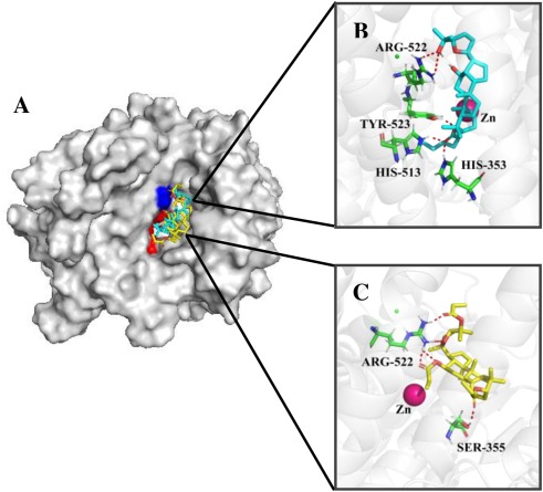
Fig. 5 Fig. 5. (A) Docking poses for compound 2e in cyan, 3b in yellow, on ACE (PDB: 1O8A). Identical residues were shown in blue and different residues in red. (B) Binding mode of compound 2e. (C) Binding mode of compound 3b. Green sticks: residues involved in the interactions; Red dashed lines: hydrogen bonds; Magenta sphere: catalytic zinc ion. (For interpretation of the references to colour in the Figure, the reader is referred to the web version of this article).
Acknowledgments
This image is the copyrighted work of the attributed author or publisher, and
ZFIN has permission only to display this image to its users.
Additional permissions should be obtained from the applicable author or publisher of the image.
Full text @ Biomed. Pharmacother.