
|
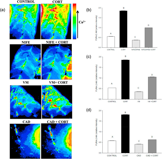
Fig. 5 Fig. 5. Cortisol-mediated [(Ca2+)i] rise involves plasma membrane Ca2+ channels. (a) Representative images of the developing trunk muscle (DTM) loaded with the ratiometric dye FURA-2AM and treated with either vehicle (CONTROL), cortisol (CORT) or the Ca2+ channel blockers nifedipine (NIFE), verapamil (VM) or cadmium (CAD) either without or with cortisol (NIFE + CORT; VM + CORT; CAD + CORT). (b) A bar graph showing the ratiometric intensity at 30 s with the L-type calcium channel inhibitor, nifedipine either with or without cortisol. (c) A bar graph showing the ratiometric intensity at 30 s with the T-type calcium inhibitor verapamil either with or without cortisol. (d) A bar graph showing the ratiometric intensity at 30 s with non-specific calcium channel inhibitor cadmium either with or without cortisol. All bars represent mean ± SEM (n = 4–5 independent embryos); Bars with different letters are significantly different; one way ANOVA; Holm Sidak post hoc test; p < 0.05.
Reprinted from Molecular and Cellular Endocrinology, 520, Das, C., Faught, E., Vijayan, M.M., Cortisol rapidly stimulates calcium waves in the developing trunk muscle of zebrafish, 111067, Copyright (2020) with permission from Elsevier. Full text @ Mol. Cell. Endocrinol.