
|
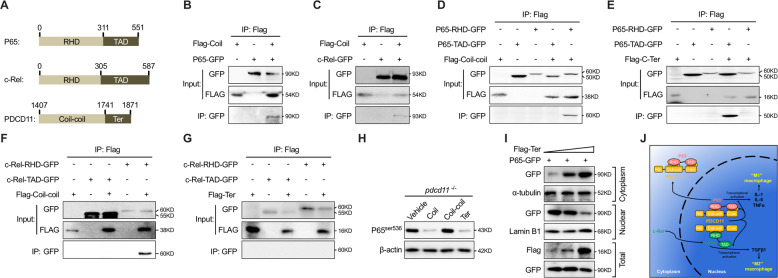
Fig. 5 a Schematic diagram showing the domain structures of human P65, c-Rel, and PDCD11-Coil. b Immunoprecipitation (IP) assay of the interaction between P65-GFP and Flag-Coil. c IP assay of the interaction between c-Rel-GFP and Flag-Coil. d IP assay of the interaction between the Coil–coil fragment of PDCD11 with the RHD domain or TAD domain of P65. e IP assay of the interaction between the terminal (Ter) fragment of PDCD11 with the RHD domain or TAD domain of P65. f IP assay of the interaction between the Coil–coil fragment of PDCD11 with the RHD domain or TAD domain of c-Rel. g IP assay of the interaction between the terminal (Ter) fragment of PDCD11 with the RHD domain or TAD domain of c-Rel. h Western blot examination of the influential role of overexpression Coil, Coil–coil and Terminal of PDCD11 on P65ser536 expression in 22 hpf pdcd11 mutants. i Western bot show the effect of increasing Ter transfection on nuclear/cytoplasm P65 content in 293T cells. j Schematic diagram showing the working model for PDCD11-mediated regulation of c-Rel and P65. Under normal circumstances, on one hand, the Ter fragment of PDCD11 (yellow) binds and masks the phosphorylation site of P65 (pink), which is required for its nuclear translocation and transcriptional activation; on the other hand, the only existing interaction between the coil–coil of PDCD11 and the RHD domain of c-Rel contrarily promotes c-Rel (green) nuclear retention for activating TGFβ1 expression, which drives macrophage differentiation toward microglia with “M2” properties. However, cells deficient in PDCD11 show increased P65 nuclear translocation, which tends to lead to activation of inflammation-related genes, such as IL-1, IL-6, and TNFα. These cytokines then facilitate “M1” macrophage differentiation.