
|
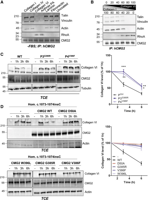
Fig. 7 Figure 7. Effect of Cytosolic HFS Mutations on Collagen VI Degradation (A) RPE1 cells were serum starved overnight and plated the next day on dishes coated with either collagen I, IV, VI, PA, laminin, fibronectin, or uncoated. After 1 h of adhesion, the cells were lysed. (B) RPE1 cells were serum starved overnight and plated the next day on varying concentrations of collagen I and collagen VI. After 1 h of adhesion, the cells were lysed. (A and B) CMG2 was immunoprecipitated (IP). IPs were then analyzed by SDS-PAGE and western blotting against talin, vinculin, actin, RhoA, and CMG2. (C and D) (C) Control, P3, and P4 fibroblasts or (D) P7 fibroblasts recomplemented with WT or HFS CBD mutants were treated with purified full-length collagen VI at 1 μg/mL. Cells were harvested 1, 3, or 6 h later. Collagen VI degradation was assessed by SDS-PAGE using 4%–12% Bis-Tris gradient gels under non-reducing conditions and western blotting for collagen VI, endogenous CMG2, and tubulin as a loading control. Right panels: the collagen VI signal was quantified by densitometric analysis. Values were normalized to the collagen VI level at 1 h. Error bars represent SEM; (C) n = 11 or (D) n = 4 for control cells, (C) n = 6 or (D) n = 4 for mutants; ∗p < 0.05; ∗∗p < 0.01; ∗∗∗p < 0.001; two-tailed unpaired t test to control.
Reprinted from Developmental Cell, 53, Bürgi, J., Abrami, L., Castanon, I., Abriata, L.A., Kunz, B., Yan, S.E., Lera, M., Unger, S., Superti-Furga, A., Peraro, M.D., Gaitan, M.G., van der Goot, F.G., Ligand Binding to the Collagen VI Receptor Triggers a Talin-to-RhoA Switch that Regulates Receptor Endocytosis, 418-430.e4, Copyright (2020) with permission from Elsevier. Full text @ Dev. Cell