
|
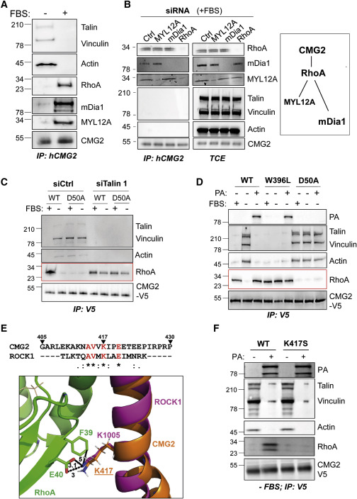
Fig. 4 Figure 4. Ligand-Bound CMG2 Interacts with RhoA, mDia1, and MYL12A (A and B) (A) RPE1 cells were treated with or without FBS overnight (B) or were transfected with control siRNA or with siRNA against MYL12A, mDia1, or RhoA for 72 h in the presence of serum. The cells were then harvested, and CMG2 was immunoprecipitated. IPs and total cell extracts (TCE) were analyzed by SDS-PAGE and western blotting against talin, vinculin, actin, RhoA, mDia1, MYL12A, and CMG2. Right panel: schematic representation of the CMG2 interaction with RhoA and associated effector proteins. (C) HeLa cells were transfected with control siRNA or with siRNA against talin 1 for 72 h, then transfected for 24 h with the cDNA expressing CMG2-V5 WT or D50A and finally serum starved overnight or not. (D) HeLa cells were transfected for 24 h with cDNA expressing CMG2-V5 WT, D50A, or W396L and grown with or without FBS overnight. The cells were then incubated or not (control) with 500 ng/mL of PA at 4°C for 1 h and switched to 37°C for 10 min before lysis. (E) Sequence alignment between residues 405 to 430 of CMG2 and the RhoA-binding domain of ROCK. One dot, two dots, and a star, respectively, indicate a semi-conservative change, a conservative change, and a perfect match. RhoA (green) and ROCK (magenta) are from PDB 1S1C, and the CMG2 segment was modeled as an ideal helix (orange, slightly kinked at a proline). The distances (in Å) are shown in black dotted lines. (F) HeLa cells were transfected for 24 h with cDNA expressing CMG2 WT or K417S, serum starved overnight and treated with 500 ng/mL of PA at 4°C for 1 h and switched to 37°C for 10 min before lysis. (C, D, and F) After cell lysis, CMG2 was immunoprecipitated. IPs were then analyzed by SDS-PAGE and western blotting against talin, vinculin, actin, RhoA, and V5. Western blots are representative of at least three independent experiments.
Reprinted from Developmental Cell, 53, Bürgi, J., Abrami, L., Castanon, I., Abriata, L.A., Kunz, B., Yan, S.E., Lera, M., Unger, S., Superti-Furga, A., Peraro, M.D., Gaitan, M.G., van der Goot, F.G., Ligand Binding to the Collagen VI Receptor Triggers a Talin-to-RhoA Switch that Regulates Receptor Endocytosis, 418-430.e4, Copyright (2020) with permission from Elsevier. Full text @ Dev. Cell