
|
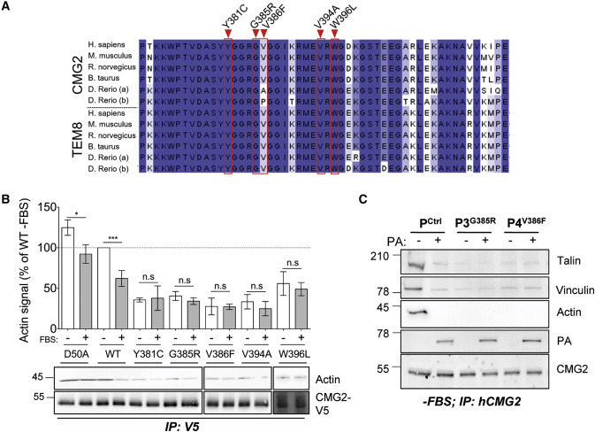
Fig. 2 Figure 2. HFS Mutations Mapping to the CMG2 Cytoplasmic Tail (A) Sequence alignment of the highly conserved cytoplasmic domain of CMG2 and TEM8. The position of the cytoplasmic HFS mutations analyzed in this study are highlighted in red. (B) Top: quantification of the interaction between CMG2 and actin by densitometry. Each value was normalized to the average signal of actin bound to WT CMG2 in cells grown in complete medium. For statistical analysis, an unpaired t test was performed on normalized values comparing the non-starved and starved condition for each cDNA construct. n = 3, ∗∗∗p < 0.001; ∗p < 0.05. Bottom: HeLa cells were transfected for 24 h with cDNA expressing CMG2-V5 WT, and mutants, grown with or without FBS overnight. The cells were lysed, and CMG2 was immunoprecipitated (IP). Samples were analyzed by SDS-PAGE and western blotting against actin and V5. (C) Control, P3, and P4 patient fibroblasts were serum starved overnight and incubated or not with 500 ng mL−1 of PA at 4°C for 1 h plus 10 min at 37°C. IPs were performed as in (A). (B and C) Representative experiments of n = 3 independent experiments.
Reprinted from Developmental Cell, 53, Bürgi, J., Abrami, L., Castanon, I., Abriata, L.A., Kunz, B., Yan, S.E., Lera, M., Unger, S., Superti-Furga, A., Peraro, M.D., Gaitan, M.G., van der Goot, F.G., Ligand Binding to the Collagen VI Receptor Triggers a Talin-to-RhoA Switch that Regulates Receptor Endocytosis, 418-430.e4, Copyright (2020) with permission from Elsevier. Full text @ Dev. Cell