
|
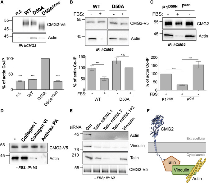
Fig. 1 Figure 1. CMG2 Interaction with the Actin Cytoskeleton (A) HeLa cells transiently transfected with cDNA expressing CMG2-V5 WT, D50A, or D50A with a deletion of the predicted CBD (D50AΔCBD) were lysed and CMG2 immunoprecipitated. IPs were then analyzed by SDS-PAGE and western blotting against actin and CMG2-V5. (B) HeLa cells were transiently transfected with cDNA expressing CMG2-V5 WT or D50A were FBS starved or not overnight, lysed and CMG2 immunoprecipitated. IPs were then analyzed by SDS-PAGE and western blotting against actin and CMG2-V5. (C) Control fibroblasts and fibroblasts from patient 1 carrying the D50N mutation. Cells were FBS starved or not overnight, lysed and CMG2 immunoprecipitated. IPs were then analyzed by SDS-PAGE and western blotting against actin and endogenous CMG2. (A–C) Actin was quantified by densitometry. Error bars represent standard error, n = 3; paired t test between D50A or D50N mutants and other constructs. n.s.: p > 0.05; ∗∗∗p < 0.001. (D) HeLa cells were transfected for 24 h with cDNA expressing CMG2 WT and plated on dishes coated with collagen I, collagen VI, or anthrax PA. After 1 h of adhesion, cells were lysed and CMG2 was immunoprecipitated. IPs were then analyzed by SDS-PAGE and western blotting against actin and CMG2-V5. (E) HeLa cells were transfected with siRNAs against talin (siRNA1 and 2), or vinculin for 72 h, and with cDNA expressing CMG2 WT for 24 h and finally serum starved overnight. Cells were then lysed, and CMG2 was immunoprecipitated. IPs were then analyzed by SDS-PAGE and western blotting against actin, vinculin, talin and CMG2-V5. (D and E) Representative experiment of n = 3. (F) Schematic representation of CMG2 interaction with the actin cytoskeleton. The cytoplasmic tail of CMG2 interacts with talin, which in turn interacts with vinculin, which is connected to actin.
Reprinted from Developmental Cell, 53, Bürgi, J., Abrami, L., Castanon, I., Abriata, L.A., Kunz, B., Yan, S.E., Lera, M., Unger, S., Superti-Furga, A., Peraro, M.D., Gaitan, M.G., van der Goot, F.G., Ligand Binding to the Collagen VI Receptor Triggers a Talin-to-RhoA Switch that Regulates Receptor Endocytosis, 418-430.e4, Copyright (2020) with permission from Elsevier. Full text @ Dev. Cell