
|
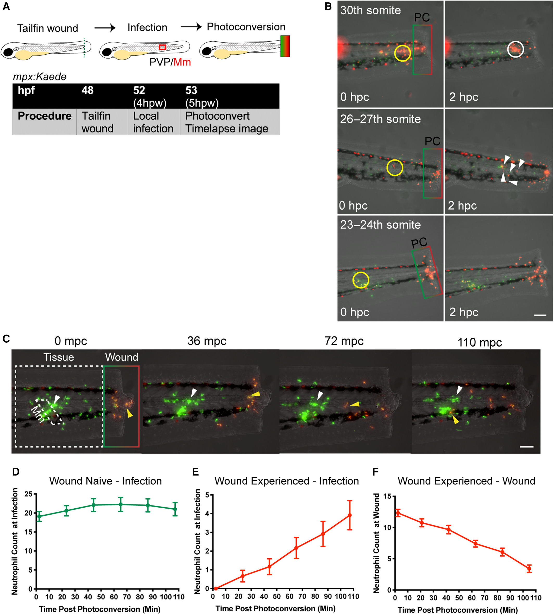
Fig. 5 Neutrophils preferentially migrated to a new infection stimulus rather than patrol the wound site. (A) Schematic of experiment for B–F. (B) Stereo‐fluorescence micrographs of a tailfin transected mpx:Kaede embryo after either 30th somite, 26/27th somite, or 23–24 somite infection with Mm at 0 and 2hpi. The local infection site is shown by a yellow ring, and PC of wound neutrophils is shown by the box. With 30 somite injection red (wound‐experienced) neutrophils have started migration to the infection site by the time the time lapse has been started and are almost all at the infection site by 2 hpi (white ring). The 26‐27th somite infection does not start recruiting wound‐experienced neutrophil by the time of the time lapse, but has done so after 2 hpi (white arrowheads). Infection into the 23rd–24th somite does not recruit any wound‐experienced neutrophils over a 2‐h time course. Representative example from n = 9 embryos per group from three independent experiments. Scale bar = 150 μm. (C) Stereo‐fluorescence micrographs of a tailfin transected mpx:Kaede embryo after 26/27th somite infection with Mm. Wound‐naïve neutrophils are green only, and those photoconverted at the wound at timepoint zero (wound‐experienced) begin as red‐only and regain GFP (therefore giving a yellow overlay) over the course of the time lapse as nascent Kaede fluorescent protein is made. Both wound‐naïve (white arrowhead) and wound‐experienced (yellow arrowhead) are recruited to the localised site of Mm infection before 110 mpc, even though the time lapse is begun at 5 hpw, a timepoint when neutrophils would normally still be recruited to the tailfin transection. Representative example from data shown in D‐F, n = 12 embryos from three independent experiments. Scale bar = 100 μm. (D) Number of green, wound‐naïve neutrophils at infection site over 1.5 hpi. Data shown are mean ± SEM, n = 12 embryos accumulated from three independent experiments. (E) Number of red, wound‐experienced neutrophils at infection site over 1.5 hpi. Data shown are mean ± SEM, n = 12 embryos accumulated from three independent experiments. (F) Number of red, wound‐experienced neutrophils at wound site over 1.5 hpi. Data shown are mean ± SEM, n = 12 embryos accumulated from three independent experiments.