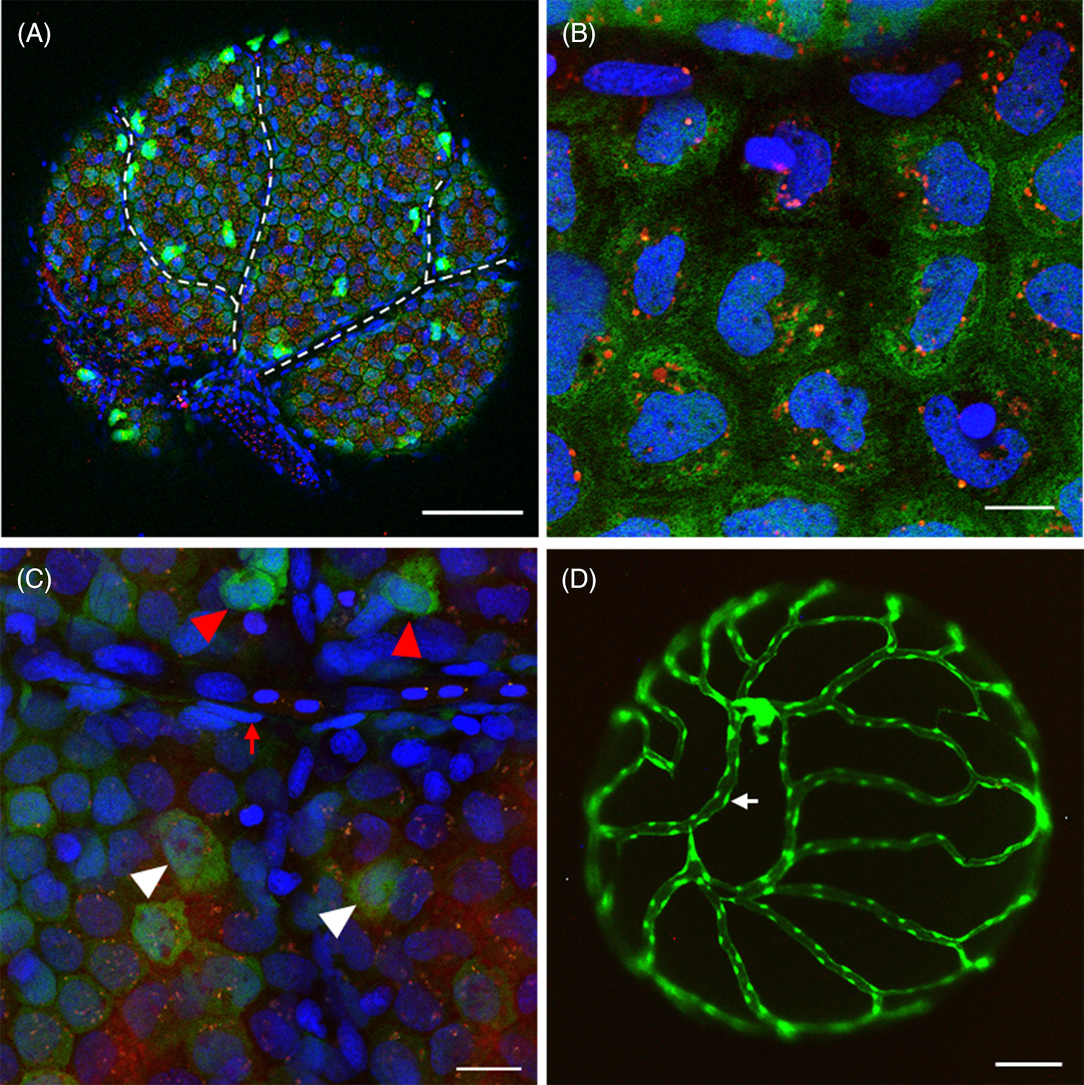
Fig. 6 Direct observation of the surface of intact ovarian follicles in Tg(pgr:egfp/gsdf:nfsB‐mCherry) and Tg(fli:egfp). A, Direct observation of the intact ovarian follicle showed almost all granulosa cells expressing both mCherry and green fluorescent protein (GFP) signals, and a few cells exhibiting strong GFP signals. The dash line indicated a network of interconnected, unstained (dark) channels without cells presenting. B, Granulosa cells expressed both mCherry and GFP signals. Noted mCherry signals present as a few spots inside granulosa cells. C, Non‐granulosa cells only exhibited strong GFP signals (red arrow head) or without GFP/mCherry signals (red arrow). White arrow head indicated granulosa cells. D, Blood vessel nest on the surface of the ovarian follicle in Tg(fli:egfp). The white arrow indicated the vascular endothelial cell. The blue fluorescence represented the nucleus which was stained by Hoechst33342. Scale bars = 100 μm (A, D), 20 μm, C, and10 μm, B. Pgr, progesterone receptor
Image
Figure Caption
Acknowledgments
This image is the copyrighted work of the attributed author or publisher, and
ZFIN has permission only to display this image to its users.
Additional permissions should be obtained from the applicable author or publisher of the image.
Full text @ Dev. Dyn.

