
|
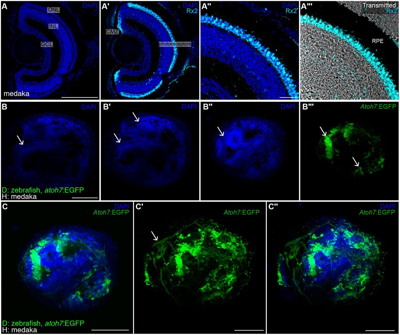
Fig. 4 Topological organisation of retinal ganglion cells in ectopic retina. (A-A‴) DAPI and immunostaining using an anti-Rx2 antibody on a medaka retina cryosection. Retinal ganglion cells localise to the ganglion cell layer (GCL) and Rx2+ photoreceptors (cyan staining in A′-A‴) are found in the outer nuclear layer (ONL), adjacent to the retinal pigmented epithelium (black region in the transmitted channel, A‴). (B-C″) DAPI staining of whole-mount retinae in zebrakas shows conspicuous layering (arrows in B,B′) and clusters of retinal ganglion cells (arrows in B″,B‴) labelled in green using a Tg(atoh7:EGFP) zebrafish donor. Single plane (C) and stack (C′,C″) showing RGCs and their axons (arrow in C′) in an ectopic zebraka retina (n=6 retinae in six chimeras). Scale bars: 100 µm in A,A′,B-C″; 20 µm in A″,A‴. GCL, ganglion cell layer; INL, inner nuclear cell layer; ONL, outer nuclear cell layer; CMZ, ciliary marginal zone; RPE, retinal pigmented epithelium; D, donor; H, host.