IMAGE

FIG. 3.

ID
ZDB-IMAGE-210113-94
Source
Figures for Cheng et al., 2020
Image
Figure Caption

FIG. 3.

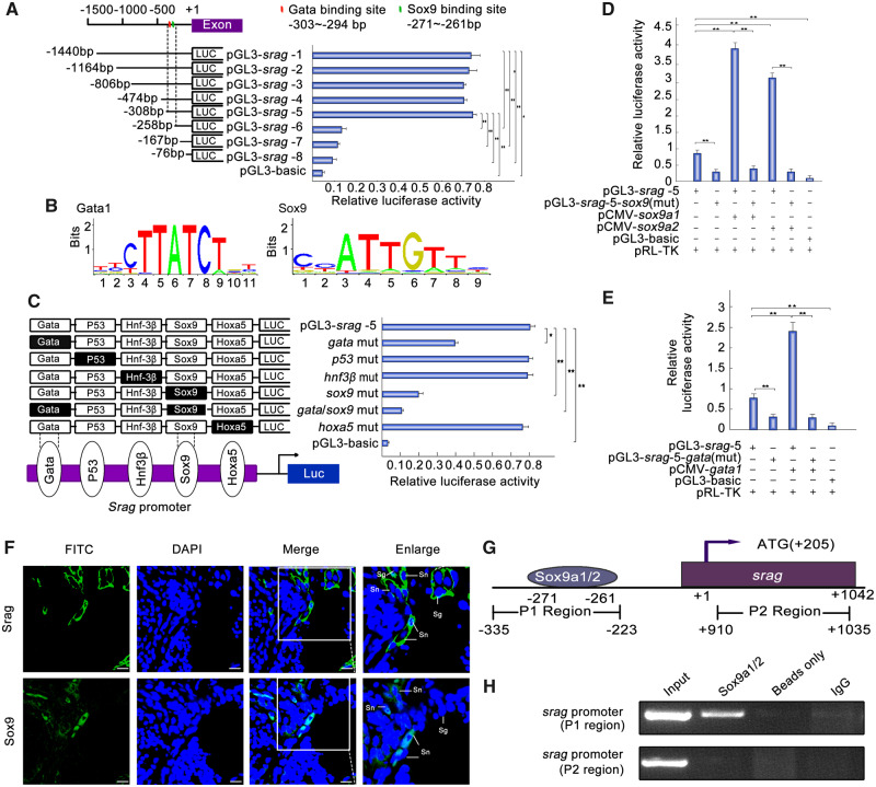
Sox9a1/2 and Gata1 upregulates srag promoter activity. (A) Luciferase assay indicated the activities of a series of truncated srag promoters in 293T cells. Endogenous SOX9 is expressed in 293T cells. Left panel showed each truncated mutant linked with the luciferase gene in the pGL3-basic vector. Binding sites of Gata and Sox9 were indicated in red and green bars, respectively. Right panel indicated the relative activities of these constructs, as determined by luciferase assays. (B) Sequence logo of Sox9- and Gata1-binding sites based on JASPAR database. (C) Point mutation analysis of the promoter using luciferase assays. The pGL3-srag-5 of 308 bp was used as a template for the point mutation analysis. Luciferase assays were used to determine the relative activities. The intact binding sites of the Gata, P53, Hnf3β, Sox9, and Hoxa5 are indicated by open boxes, respectively. The filled boxes show the corresponding mutations. The pGL3-basic vector was used as a negative control. (D) Sox9a1/2 overexpression upregulated the luciferase activity of srag promoter. Sox9a1/2 transfection activated the srag promoter. In total, 0.32 μg pGL3-srag-5 or its Sox9-binding site mutant (pGL3-srag-5-sox9(mut)) was cotransfected with 0.08 μg sox9a1/2 expression plasmids (pCMV-sox9a1 and pCMV-sox9a2), as indicated. Sox9a1 and Sox9a2 overexpression increased the activity of pGL3-srag-5 but did not affect the activity of pGL3-srag-5-sox9(mut). (E) Gata1 overexpression upregulated the luciferase activity of srag promoter. Gata1 transfection activated the srag promoter. In total, 0.32 μg pGL3-srag-5 or its Gata-binding site mutant (pGL3-srag-5-gata (mut)) was cotransfected with 0.08 μg Gata1 expression plasmids (pCMV-gata1). Gata1 overexpression increased the activity of pGL3-srag-5, but did not affect the activity of pGL3-srag-5-gata (mut). (F) Coexpression analysis between Srag and Sox9. Immunofluorescence of testis samples in serial sections using anti-Srag or anti-Sox9 antibody followed by FITC-conjugated ImmunoPure goat anti-rabbit IgG (green). Sox9 was expressed in the nuclei of Sertoli cells (Sn) in testis, and Srag was detected mainly in the cytoplasm of Sertoli cells (Sn) and spermatogonia (Sg). The nuclei were stained by DAPI (blue). Images were captured using confocal microscopy. The enlarged images originated from the regions with white squares. Scale bar: 10 μm. (G) Schematic diagram of primer relative positions in the ChIP assays. (H) ChIP assays. ChIP analysis showed that Sox9 could bind to the srag promoter in vivo. Sonicated chromatin from testis of the adult individuals was used for PCR amplification. A 112-bp fragment corresponding to the −335 to −223 region of the srag promoter was amplified using the immunoprecipitated DNA as a template which was immunoprecipitated with monoclonal anti-Sox9. In controls (no antibody [beads only] or preimmune IgG), no band from the anti-Sox9 antibody precipitates was observed. Exon of srag was used as a negative control. The means ± SD are from three independent experiments. One-way ANOVA was performed. *P < 0.05; **P < 0.01.

Acknowledgments
This image is the copyrighted work of the attributed author or publisher, and ZFIN has permission only to display this image to its users. Additional permissions should be obtained from the applicable author or publisher of the image. Full text @ Mol Bio Evol