IMAGE

FIGURE 5

ID
ZDB-IMAGE-210113-31
Genes
Source
Figures for Mignani et al., 2020
Image
Figure Caption

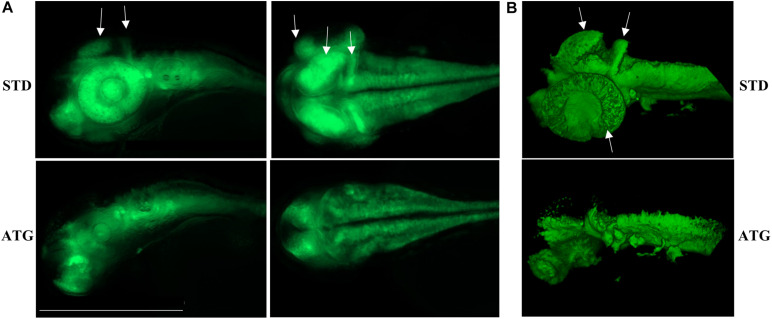
FIGURE 5

neurod1-dependent EGFP fluorescence in embryos injected with control (STD) or ATG morpholinos. (A) Representative lateral and dorsal views of Tg(neurod1:EGFP) embryos either injected with STD or ATG morpholinos, at 48 hpf, Size bar = 500 μm. Experiments were repeated at least three times and altogether more than 150 embryos of each type were analyzed. (B) Lightsheet imaging of embryos described in panel (A). Four embryos with mild morphological phenotype were randomly selected and analyzed in two different experiments. Arrows point to the regions that are clearly missing or less visible in ATG-morphants.

Figure Data
Acknowledgments
This image is the copyrighted work of the attributed author or publisher, and ZFIN has permission only to display this image to its users. Additional permissions should be obtained from the applicable author or publisher of the image. Full text @ Front Cell Dev Biol