IMAGE

Fig. 2

ID
ZDB-IMAGE-201210-8
Source
Figures for Everson et al., 2020
Image
Figure Caption

Fig. 2

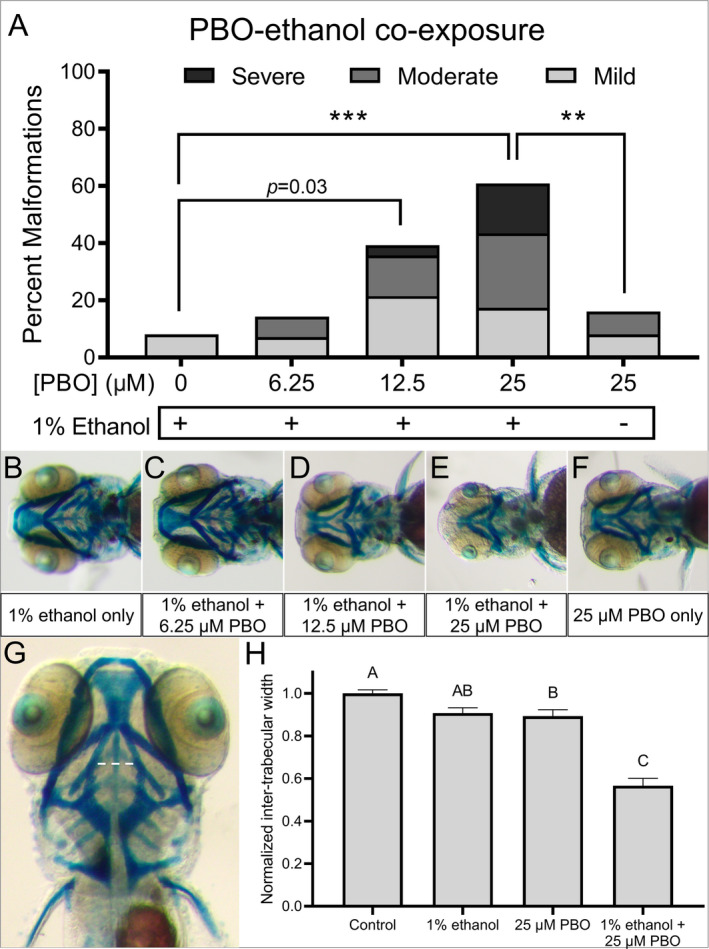
Ethanol and PBO synergistically interact to cause craniofacial defects. (A) Wild‐type embryos were exposed to 0, 6.25, 12.5, or 25 μM PBO with or without a 1% dose of ethanol. Percent malformations (mild, moderate, and severe) are shown. Incidence of malformations was compared between treatment groups using Fisher’s exact test with Bonferroni correction for multiple comparisons. ***p < 0.001, **p < 0.01. (BF) Whole‐mount images of alcian‐ and alizarin‐stained embryos are shown for each treatment group. (G) 5‐day‐old alcian‐ and alizarin‐stained embryo marks the measurement for inter‐trabecular width (white dashed outline). (H) Quantification of inter‐trabecular widths. Mean width ± SEM is shown. Measurements were normalized to control and compared using one‐way ANOVA with Tukey’s multiple comparisons test. Different letters indicate statistically significant differences between the groups. See supplemental data for the specific multiple comparisons p‐values. N ≥ 28 embryos per treatment.

Figure Data
Acknowledgments
This image is the copyrighted work of the attributed author or publisher, and ZFIN has permission only to display this image to its users. Additional permissions should be obtained from the applicable author or publisher of the image. Full text @ Alcoholism Clin. Exp. Res.