IMAGE

FIGURE 2

ID
ZDB-IMAGE-201121-42
Source
Figures for Kuroda et al., 2020
Image
Figure Caption

FIGURE 2

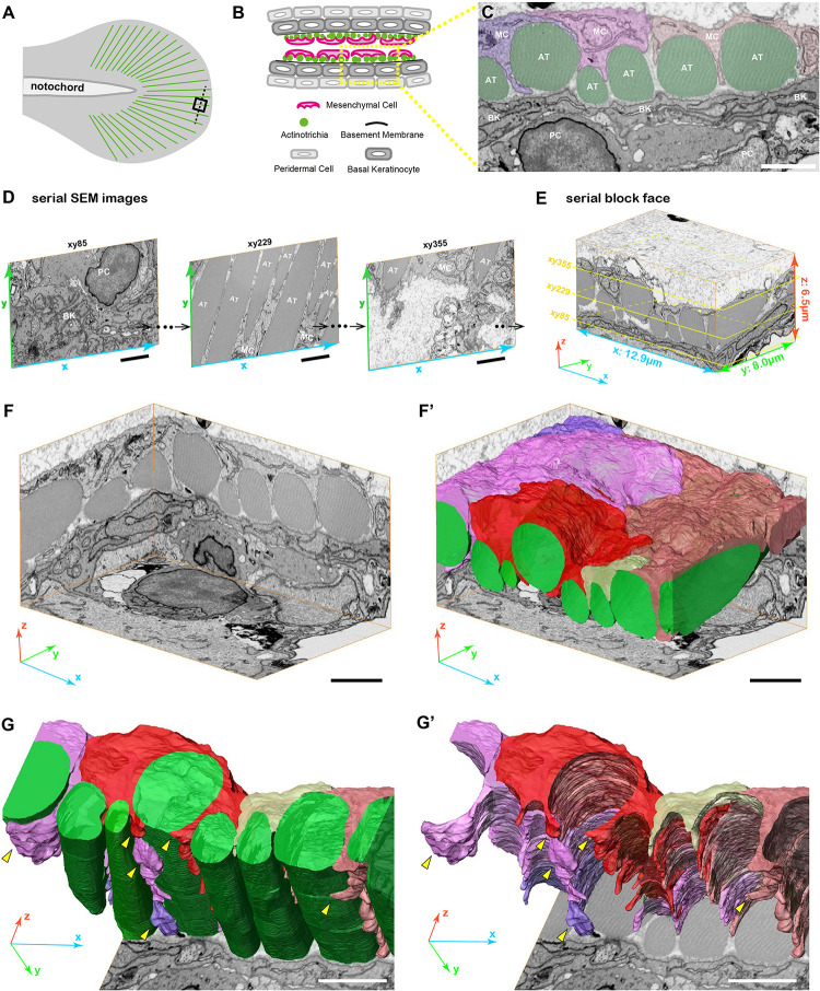
3D reconstruction of fin mesenchymal cells and actinotrichia by FIB-SEM analysis. (A) Illustration of the 3 dpf larval median fin. Black boxed region (15 μm × 15 μm) in the fin tip was used for the FIB-SEM analysis. (B) Schematic diagram of the transverse section of the black dotted line in (A) at the fin tip. Actinotrichia are distributed inside the basement membrane. (C) An example of SEM image of the transverse section at the fin tip. Mesenchymal cells (shown in purple, pink, and orange color) develop the long pseudopodia structures and directly interact with actinotrichia (shown in green color). (D) Serial SEM images at x-y- plane obtained by FIB-SEM observation. (E) Three-dimensional view of the serial block surface image, 12.9 μm × 8.0 μm × 6.5 μm. The positions of each SEM images in (D) are shown in the yellow boxes. (F) Three-dimensional view of the reconstructed sequential SEM images and (F′) the 3D reconstruction of eight actinotrichia (labeled by green color) and five mesenchymal cell domains (labeled by purple, pink, red, yellow, and orange color). (G) 3D reconstruction of actinotrichia and mesenchymal cell domains and (G′) 3D reconstruction of mesenchymal cell domains viewed from the epidermal cell layer. Multiple pseudopodia structures are developed from each mesenchymal cell domains and hold actinotrichia fibrils (yellow arrowheads). MC, mesenchymal cell; AT, actinotrichia; BK, basal keratinocyte; PC, peridermal cell. Scale bars: 2 μm.

Acknowledgments
This image is the copyrighted work of the attributed author or publisher, and ZFIN has permission only to display this image to its users. Additional permissions should be obtained from the applicable author or publisher of the image. Full text @ Front Cell Dev Biol