
|
Fig. 4
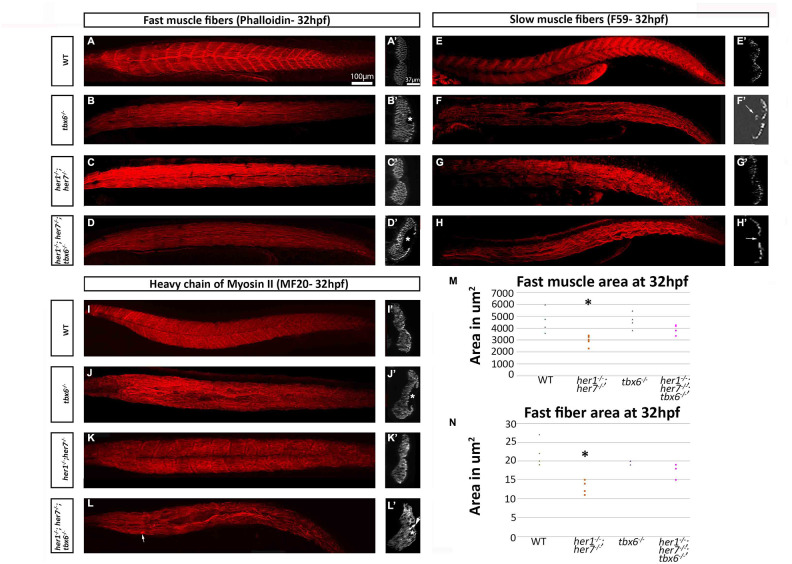
Fast and slow muscle fibers are affected in mutants at embryonic stages. (A–D) Phalloidin staining for fast muscle fibers in all mutants show loss of the characteristic metameric structure. (A) In addition, the transverse plane sections at the cloacal level in tbx6-/- and her1-/-; her7-/-; tbx6-/- display hollow cavities where no muscle fibers were present (B´ and D´). F59 staining for slow muscle fibers (E–H) also demonstrates loss of the characteristic metameric patterning in the mutants. tbx6-/- and her1-/-; her7-/-; tbx6-/- mutants present fusions of the fibers and cavities (F, F´ and H´). The her1-/-; her7-/- slow fibers resemble wild type fibers in sections (G´). The marker for striated muscle, MF20 (I–L) shows the same phenotype as phalloidin and allows the visualisation of aggregates of the fibers near the areas where there are lesions in the her1-/-; her7-/-; tbx6-/- mutants (arrow in L´). Quantifications of fast muscle area (M) and fiber cross-sectional area (N) at 32hpf display a statistically significant decrease (p= 0.03 and p= 0.009 respectively) of these two criteria in the her1-/-; her7-/ - mutants. In both figures, 6 points were measured. An asterisk denotes when the difference between the wild types and mutants is statistically different (p<0.05).