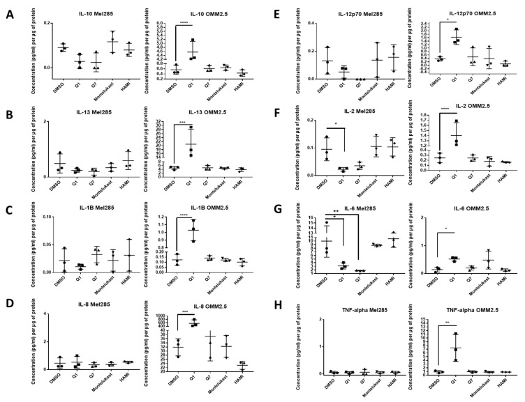
IMAGE

Figure 7

ID
ZDB-IMAGE-201102-34
Source
Figures for Slater et al., 2020
Image
Figure Caption

Figure 7

ELISA of cell conditioned media demonstrates that 24 h treatment with 20 µM quininib analogues decreases inflammatory markers in Mel285 cells but increases inflammatory markers in OMM2.5 cells. In OMM2.5 cells, 24 h treatment with quininib (Q1) significantly increased the secretion of IL-10 (A), IL-13 (B), IL-1B (C), IL-8 (D), IL-12p70 (E), IL-2 (F), IL-6 (G), and TNF-α (H). In Mel285 cells, 24-h treatment with 20 μM quininib (Q1) and 1,4–dihydroxy quininib (Q7) significantly reduced the levels of IL-2 (F) and IL-6 (G). Treatment with 20 μM montelukast or HAMI 3379 had no effect on the secretion of inflammatory markers in either cell line. Conditioned media were collected from three separate experiments and analysed by ELISA (n = 3). All secretions were normalised to total protein content. Statistical analysis was performed by ANOVA with Dunnett’s post hoc multiple comparison test. Error bars are mean + S.E. * p < 0.05; ** p < 0.01; *** p < 0.001; **** p < 0.0001.

Acknowledgments
This image is the copyrighted work of the attributed author or publisher, and ZFIN has permission only to display this image to its users. Additional permissions should be obtained from the applicable author or publisher of the image. Full text @ Cancers