
Fig. 1
|
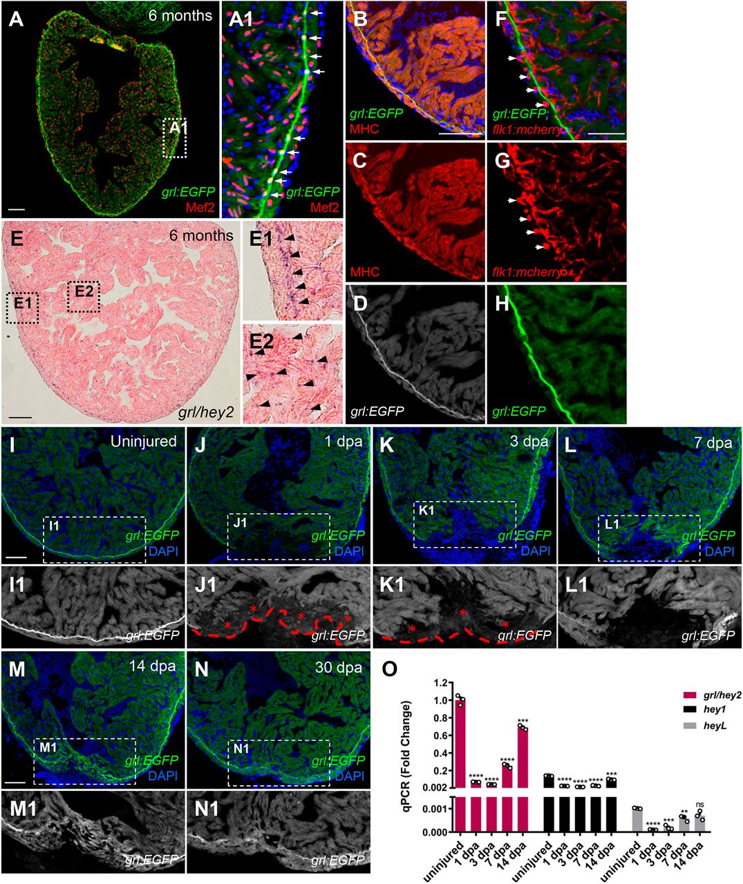
Fig. 1 Reduction of myocardial grl correlates with regenerative responses of the zebrafish heart to injury. (A-D) grl:EGFP (green or white) overlaps with a nuclear CM marker Mef2 (red) (A) and a cytoplasmic CM marker MHC (red) (B-D). (A1) Enlarged image of the dashed box in A. Arrows indicate the grl-enriched primordial layer (PML). (E) ISH for grl displays enriched expression in the PML and its expression throughout the myocardium in adult zebrafish hearts. (E1) Higher-magnification image of the dashed box in E; arrowheads point to the grl-enriched PML. (E2) Enlarged image of the dashed box in E; arrowheads point to grl expression in the myocardium. (F-H) grl:EGFP (green) does not colocalize with endocardial or coronary endothelial cells marked by flk1:mCherry (red) in adult Tg(grl:EGFP;flk1:mCherry) hearts. Arrows indicate the circular coronary vessels. (I-N) Tg(grl:EGFP) adult heart show grl:EGFP (green) expression in uninjured and regenerating ventricles. (I1-N1) Higher-magnification images of the dashed boxes in I-N. Red dashed line indicates approximate plane of resection. Red asterisks mark decreased expression of grl:EGFP in the injury border zone. (O) Expression of grl, hey1, and heyL were examined using qPCR analyses in uninjured and regeneration ventricular samples. Expression levels were normalized to that of β-actin and further normalized to that of grl in uninjured sample (n=3). Data presents as mean±s.e.m. **P<0.01, ***P<0.001, ****P<0.0001, Student's t-test (unpaired, two-tailed). Scale bars: 100 µm (A-E, I-N); 50 µm (F-H).