IMAGE

FIGURE 2

ID
ZDB-IMAGE-201007-15
Source
Figures for Wang et al., 2020
Image
Figure Caption

FIGURE 2

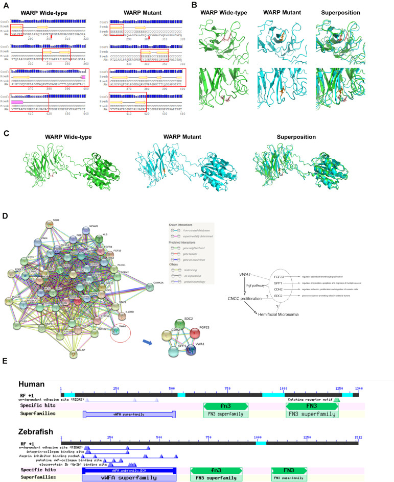
Predicted WARP structures, protein interactions and conserved domains. (A) Predicted secondary structures of wild-type and mutant protein sequences flanking the mutation. Secondary structural features are annotated as follows: pink cylinder, α-helix; yellow arrow, β-sheet; black line, coil; Conf, confidence; Pred, predicted; H in Pred line, helix; C in Pred line, coil; E in Pred line, sheet; AA, amino acid; red arrow, mutant amino acid. Local (B) and global (C) views of the predicted tertiary structures of wild-type (green) and mutant (blue) proteins. Structural superposition analyses demonstrating the change in protein structure brought about by the mutation are also displayed. (D) Protein-to-protein interactions of WARP with the FGF pathway. Predictions using the web-based program, String, indicated that WARP may functionally interact with four proteins of FGF pathway: FGF23, SPP1, CDH2, and SDC2. (E) Domains of WARP conserved in humans and zebrafish. Humans and zebrafish have the same WARP conserved domains: a von Willebrand factor A-domain, the first fibronectin type III repeat, and the second fibronectin type III repeat.

Acknowledgments
This image is the copyrighted work of the attributed author or publisher, and ZFIN has permission only to display this image to its users. Additional permissions should be obtained from the applicable author or publisher of the image. Full text @ Front Cell Dev Biol