IMAGE

Figure 3

ID
ZDB-IMAGE-201003-252
Source
Figures for Wisniewski et al., 2020
Image
Figure Caption

Figure 3

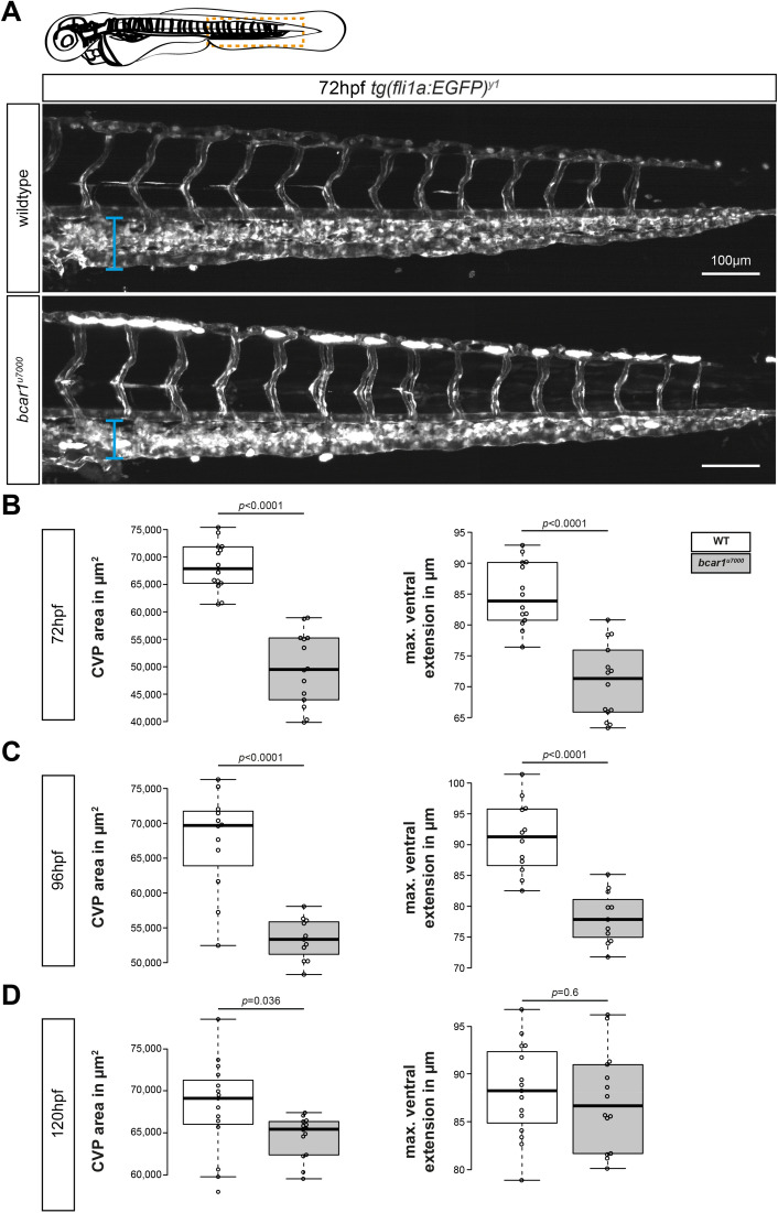
P130Cas deletion results in prolonged reduction of caudal vein plexus area. (A) Maximum intensity projections of the caudal vasculature of WT and bcar1u7000 embryos at 72hpf, from the anal pore, as illustrated by orange box in cartoon of embryo at the top. (B–D) Quantification of the maximum ventral extension of the CVP to the dorsal aorta, as well as overall vascular CVP area confirm that significant reductions remain in mutant embryos at 3dpf (B) and 4dpf (C). At 5dpf, the caudal area is still smaller but the ventral extension no longer significantly different (D). All results displayed as box plots with centre line showing median and box dimensions indicating 25th and 75th quartile. Each data point represents an individual embryo, experimental n from ≥ 2 independent clutches, p values as shown. White boxes are wildtype, light grey boxes indicate bcar1u7000. Scale bar as indicated.

Figure Data
Acknowledgments
This image is the copyrighted work of the attributed author or publisher, and ZFIN has permission only to display this image to its users. Additional permissions should be obtained from the applicable author or publisher of the image. Full text @ Sci. Rep.