
|
Fig. 4-S1
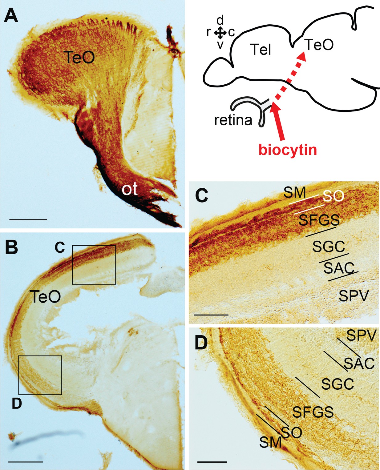
Retinal projections terminating in TeO.
(A–D) Frontal sections of the adult TeO (lateral to the left) visualizing biocytin-labeled structures subsequent injection in the optic nerve. (A) The most anterior part of TeO shown with intensively labeled optic tract (ot). (B) More posterior TeO showing the biocytin-labeled nerve terminals in the upper layer of TeO. C and D show a higher magnification of the squared areas in (B), showing the layered cytoarchitecture of the TeO. Abbreviations: ot, optic tract; SAC, stratum album centrale; SFGS, stratum fibrosum et griseum superficiale; SGC, stratum griseum centrale; SM, stratum marginale; SO, stratum opticum; SPV, stratum periventriculare; Tel, telencephalon; TeO, optic tectum. Brain orientation: r, rostral; c, caudal; d, dorsal; v, ventral. Scale bars = 200 µm (A and B); 50 µm (C and D).