
|
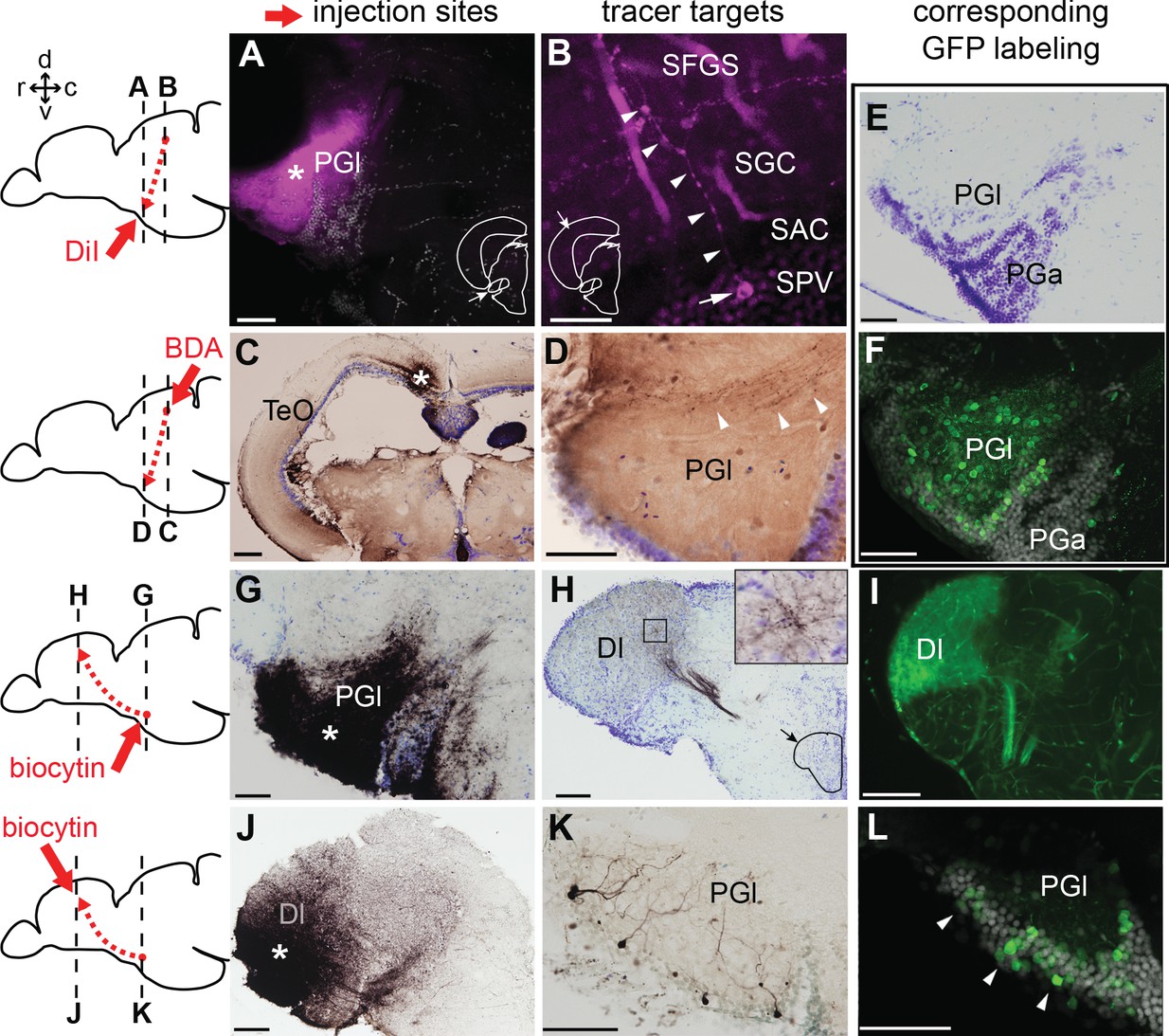
Fig. 4
Tract-tracing study showing connections of the lateral preglomerular nucleus (PGl).
Schematic drawings indicate the injection site of each tracer and levels of the frontal sections shown on the right panels. In all the frontal sections shown in A-L, the lateral side of the brain is to the left. (A–F) PGl receiving tectal inputs. (A and B) DiI retrograde labeling (magenta), showing the injection site in the PGl (A; asterisk), and a retrogradely labeled neuron (B; arrow) located in a deep layer (SPV) of the optic tectum (TeO). This neuron extends its dendrites (B; arrowheads) to the upper layer (SFGS) where retinal projections terminate. (C and D) BDA anterograde labeling (brown) showing the injection site in TeO (C; asterisk) and anterogradely labeled terminals in the ipsilateral PGl (D; arrowheads). (E) Cresyl violet staining showing the cytoarchitecture of the zebrafish PG: the lateral (PGl) and the anterior (PGa) subdivisions can be identified, based on the comparison with the goldfish PG (the nomenclature applied from goldfish; Yamamoto and Ito, 2008). (F) GFP+ labeling in the PG of Tg(279A-GFP) zebrafish line (20 µm projection of confocal images; GFP in green and DAPI in grey), showing a section comparable to the level shown in (E). GFP+ perikarya are mostly found in PGl. (G–L) PGl neurons projecting to Dl of the pallium. (G and H) Biocytin injection site in the PGl (G; asterisk) and anterogradely labeled terminals in the ipsilateral Dl (H). The right top inset in (H) shows a higher magnification of the squared area showing numerous labeled terminals. (I) GFP+ fiber labeling in the Dl of Tg(279A-GFP) zebrafish line, demonstrating an arborization pattern comparable to the anterograde biocytin labeling shown in (H). (J and K) Biocytin injection site in the Dl (J; asterisk), and retrogradely labeled neurons in the ipsilateral PGl (K) that extend dendrites ramifying in the neuropil. (L) GFP+ perikarya labeling in the PGl of Tg(279A-GFP) zebrafish line (arrowheads; 5 µm projection), demonstrating the almost identical cell localization as shown in (K). Abbreviations: Dl, lateral part of dorsal telencephalic area; PGa, anterior preglomerular nucleus; PGl, lateral preglomerular nucleus; SAC, stratum album centrale; SFGS, stratum fibrosum et griseum superficiale; SGC, stratum griseum centrale; SPV, stratum periventriculare; TeO, optic tectum. Brain orientation: r, rostral; c, caudal; d, dorsal; v, ventral. Scale bars = 50 µm (A, D-G, K, and L); 20 µm (B); 200 µm (C); 100 µm (H–J).