
|
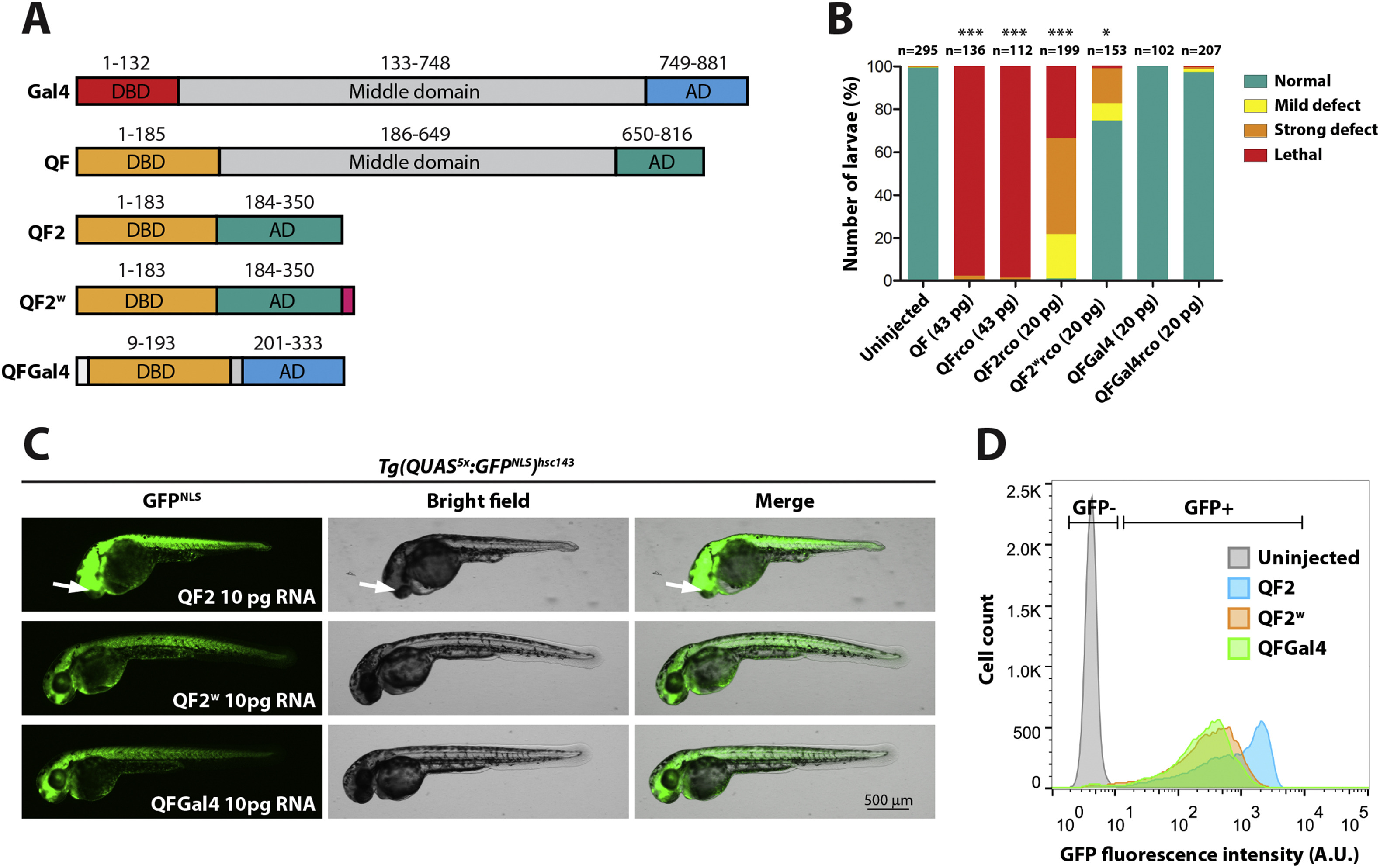
Fig. 1 QFGal4 is a non-toxic transcriptional activator in zebrafish. (A) Schematic of Gal4 (S. cerevisiae) and QF (N. crassa) protein domains. QF and Gal4 have a similar domain organization, with an N-terminal DBD, a middle domain, and a C-terminal AD. QF2 lacks the middle domain of QF, and QF2w contains an additional C-terminal polylysine tract to attenuate activity (Riabinina et al., 2015). QFGal4 contains the DNA-binding domain of QF (residues 1–185) fused to the transcriptional activation domain of Gal4 (749–881). QFGal4 also contains an SV40 NLS (PKKKRK) at the N-terminus to facilitate localization to the nucleus. (B) Bar graph showing the relative toxicity of QF and its derivatives in zebrafish embryos. Equimolar amounts of mRNA encoding QF variants were injected into one-cell stage embryos. Full length QF, both original codon usage (n = 136) and recodonized (rco) versions (n = 112), was strongly toxic with most embryos exhibiting lethality during the blastula stage (98% and 98%, respectively). QF2 recodonized (n = 199) was also toxic, with the majority of embryos exhibiting either lethality (35%) or strong developmental defects (44%). QF2w recodonized (n = 153) was well tolerated with 71% of embryos appearing healthy. QFGal4, both original codon usage (n = 102) and QF recodonized (n = 207), was well tolerated (100% and 98% normal, respectively). Injected embryos were scored as follows: mild defects included heart edema and/or mild tail kinking; severe defects included gross developmental defects, such as failure to form embryonic structures like the eye; and lethality was scored as embryos that did not proceed past the blastula stage. QF, QFrco, QF2rco and QF2wrco were significantly different from uninjected control embryos, while no significant difference was detected for either QFGal4 or QFGal4rco as compared to uninjected controls. (C) Epifluorescence micrographs showing the in vivo activity of either QF2, QF2w or QFGal4 as determined by RNA injection into a fourth-generation Tg(QUAS5x:GFPNLS)hsc143 reporter line. 10 pg of mRNA encoding either QF2 (top panel), QF2w (middle panel) or QFGal4 (lower panel) was injected at the one-cell stage and larvae were imaged at 2 dpf. Tg(QUAS5x:GFPNLS)hsc143 larvae injected with QF2 mRNA exhibited strong, ubiquitous GFPNLS expression, although larvae exhibited developmental defects, including misplaced eyes (arrow). Tg(QUAS5x:GFPNLS)hsc143 larvae injected with either QF2w or QFGal4 mRNA also exhibited ubiquitous GFPNLS expression, although signal appeared weaker than embryos injected with QF2 mRNA. (D) Plot showing relative GFPNLS fluorescence intensity of 2 dpf disassociated Tg(QUAS5x:GFPNLS)hsc143 transgenic embryos injected with 10 pg mRNA encoding either QF2, QF2w or QFGal4 mRNA. QF2 (blue) resulted in the strongest activation of a Tg(QUAS5x:GFPNLS)hsc143 reporter, followed by QF2w (orange) and QFGal4 (green). 30,000 events per sample are shown.
Reprinted from Developmental Biology, 465(2), Burgess, J., Burrows, J.T., Sadhak, R., Chiang, S., Weiss, A., D'Amata, C., Molinaro, A.M., Zhu, S., Long, M., Hu, C., Krause, H.M., Pearson, B.J., An optimized QF-binary expression system for use in zebrafish, 144-156, Copyright (2020) with permission from Elsevier. Full text @ Dev. Biol.