
IMAGE
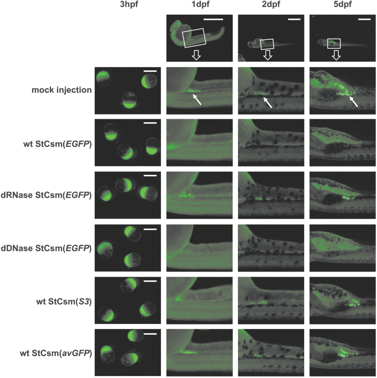
FIG. 2.
- ID
- ZDB-IMAGE-200905-8
- Publication
- Fricke et al., 2020 - Targeted RNA Knockdown by a Type III CRISPR-Cas Complex in Zebrafish
- All Figures
- Figures for Fricke et al., 2020
Image
|
Figure Caption
FIG. 2.
Microscopy of StCsm mediated
Acknowledgments
This image is the copyrighted work of the attributed author or publisher, and
ZFIN has permission only to display this image to its users.
Additional permissions should be obtained from the applicable author or publisher of the image.
Full text @ CRISPR J