IMAGE

FIG. 3.

ID
ZDB-IMAGE-200905-10
Source
Figures for Fricke et al., 2020
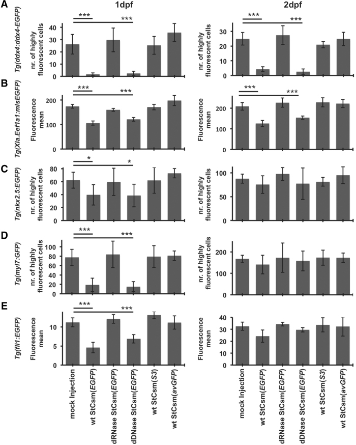
Image
Figure Caption

FIG. 3.

Quantification of EGFP knockdown efficiency by fluorescence-activated cell sorting analysis. Enhanced green fluorescent protein fluorescence in (A)Tg(ddx4:ddx4-EGFP), (B)Tg(Xla.Eef1a1:mlsEGFP), (C)Tg(nkx2.5:EGFP), (D)Tg(myl7:GFP) and (E)Tg(fli1:EGFP) fish was quantified 1 dpf and 2 dpf by flow cytometry of minced and trypsinized embryos. For Tg(ddx4:ddx4-EGFP), Tg(nkx2.5:EGFP) and Tg(myl7:GFP) embryos, the number of highly fluorescent cells (at least 50-fold background fluorescence) was counted. Results are from 3 independent experiments; error bars represent one standard deviation. *P < 0.05 as compared with respective controls, ***P < 0.001 as compared with respective controls. Data are represented as mean ± SEM.

Acknowledgments
This image is the copyrighted work of the attributed author or publisher, and ZFIN has permission only to display this image to its users. Additional permissions should be obtained from the applicable author or publisher of the image. Full text @ CRISPR J