
|
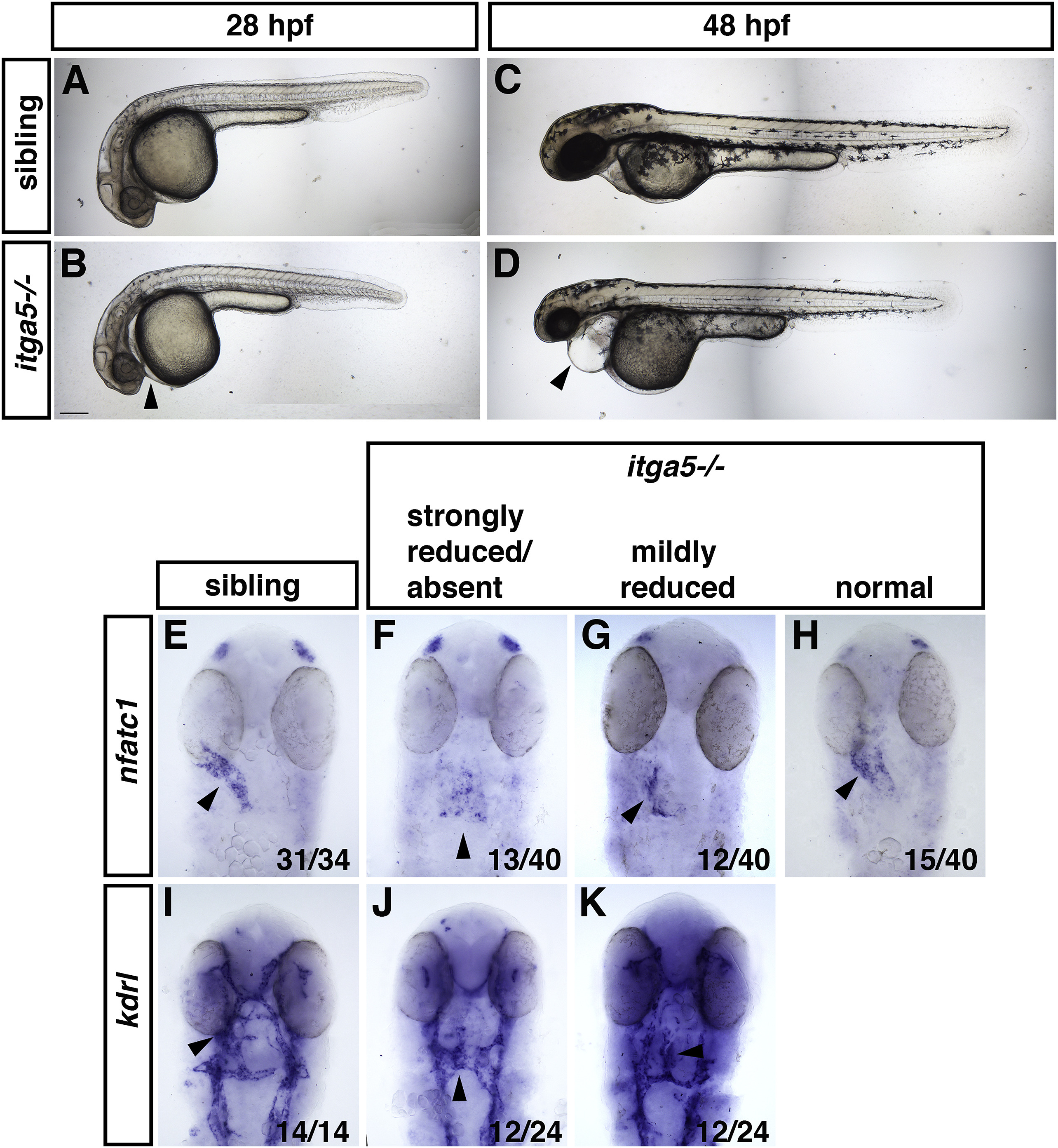
Fig. 2 Fig. 2. itga5 mutant embryos have endocardial defects. (A, B) Sibling and itga5 mutant embryos at 28 hpf. (C, D) Sibling and itga5 mutant embryos at 48 hpf. Lateral views, arrowheads indicate pericardial edema that is visible in itga5 mutants at both stages. Scale bar is 200 μm. (E–H) nfatc1 expression analyzed by ISH is reduced to varying degrees in the endocardium of itga5 mutants compared to siblings. (I–K) kdrl expression is normal in the itga5 mutant endocardium, but the morphology of the endocardium is abnormal compared to siblings. Arrowheads indicate endocardium. Embryos in E-K are at 28 hpf, dorsal views.
Reprinted from Developmental Biology, 465(1), Schumacher, J.A., Wright, Z.A., Owen, M., Bredemeier, N.O., Sumanas, S., Integrin α5 and Integrin α4 cooperate to promote endocardial differentiation and heart morphogenesis, 46-57, Copyright (2020) with permission from Elsevier. Full text @ Dev. Biol.