
|
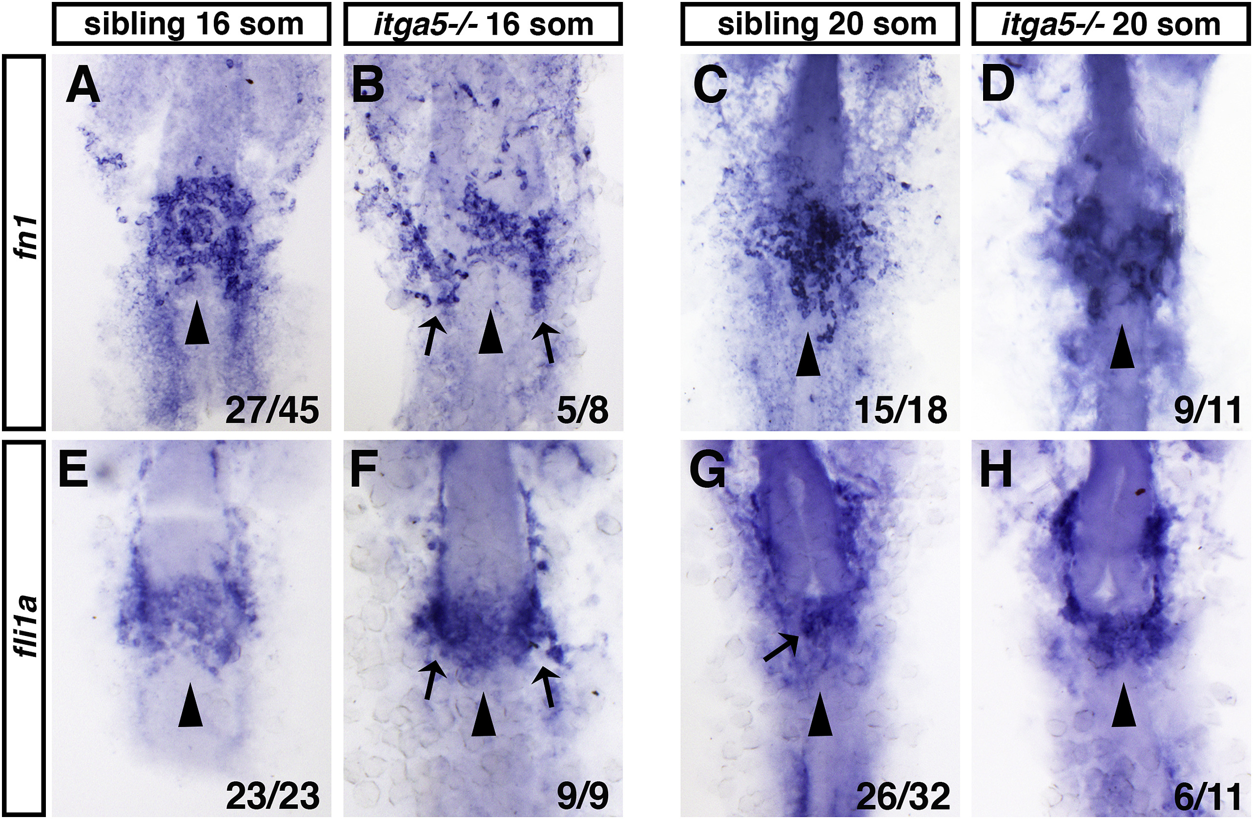
Fig. 3 Fig. 3. Endocardial specification is normal in itga5 mutants, whereas endocardial sheet formation is slightly delayed in itga5 mutants. (A–D) fn1 is expressed in endocardial progenitors of itga5 mutants at both 16 somite and 20 somite stages, but the expression domain is split or partially split. (E–H) fli1a expression in itga5 siblings and mutants at both 16 somite and 20 somite stages. Dorsal views, arrowheads indicate midline of endocardial sheet. Arrows in B and F indicate pools of endocardial progenitors that remain in more lateral locations compared to siblings. Arrow in G indicates condensation of endocardial cells in the middle of the endocardial sheet.
Reprinted from Developmental Biology, 465(1), Schumacher, J.A., Wright, Z.A., Owen, M., Bredemeier, N.O., Sumanas, S., Integrin α5 and Integrin α4 cooperate to promote endocardial differentiation and heart morphogenesis, 46-57, Copyright (2020) with permission from Elsevier. Full text @ Dev. Biol.