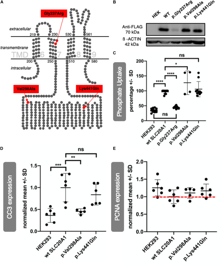
IMAGE

FIGURE 5

ID
ZDB-IMAGE-200829-28
Source
Figures for Rieke et al., 2020
Image
Figure Caption

FIGURE 5

SLC20A1 as transmembrane phosphate transporter and in vitro characterization of its variants found in BEEC individuals. (A)In silico 2D model of SLC20A1, a multi-pass integral membrane protein, indicating localization of variants found in affected individuals, c.709G > A (p.Gly237Arg) in TMD 7, c.893T > C (p.Val298Ala) and c.1321A > C (p.Lys441Gln) are located in an intracellular loop. Only four (6–9) of a total of twelve TMDs are shown in this simplified model. The model was generated based on the data of Beck et al. (2009). (B,C)SLC20A1 mediated 32PO4 transport in transiently transfected HEK293 cells. (B) WB analysis of 100 μg whole cell homogenates obtained from HEK293 cells transfected with 500 ng plasmid DNA per cm2 well surface. Plasmid DNA was FLAG tagged and transfection efficiency was detected using anti-FLAG antibody (70 kDa) and anti-ß-ACTIN antibody (42 kDa), which served as loading control. As expected, no FLAG signal could be detected in negative control (untransfected HEK293 cells, marked as HEK). Transfection worked for wt SLC20A1 overexpression as well as the variants c.893T > C (p.Val298Ala) and c.1321A > C (p.Lys441Gln). No FLAG signal could be detected for c.709G > A (p.Gly237Arg) transfected cells. Even when transfected with higher plasmid concentrations, p.Gly237Arg was not detectable in HEK293 cells (Supplementary Data Sheet S12). (C) Endpoint assay of transient transfected HEK293 cells. Cells were incubated with 1 μCi 32PO43– and 200 μM K3PO4 for 15 min. For better comparison, a highest number of counts per minute in wt SLC20A1 overexpression group was set as 100% in each experiment (N = 6 with two datasets each), values of HEK293 and variants were calculated correspondingly, and resulting values in percentage are shown on the y-axis (Error bars show SD). A two-way ANOVA of the grouped analysis was significant ((p < 0.0001). p.Gly237Arg did not show any differences of phosphate uptake to negative HEK293 control. This is in line with the expression deficiency of p.Gly237Arg described before. Wt SLC20A1 overexpression showed a significant increase of phosphate uptake compared to untransfected HEK293 (Tukey’s multiple comparison: ****p < 0.0001). Amin acid change p.Lys441Gln and p.Val298Ala overexpression resulted in an even higher phosphate uptake than wt SLC20A1 overexpression with a significant difference between p.Val298Ala and wt SLC20A1 (Tukey’s multiple comparison: *p < 0.05). Therefore, variant overexpression does not impair phosphate uptake capability in vitro. (D,E) Densitometric analysis of WBs (N = 6) from whole cell homogenates was obtained from transfected HEK293 cells. WBs are provided in Supplementary Data Sheet S13. Y-axis shows normalized values against wt SLC20A1 overexpression. Error bars show SD. (D) Expression of CC3 as apoptosis marker was measured in six WBs of corresponding independent transfection experiments. The one-way ANOVA was significant (p = 0.0002), Tukey’s multiple comparison test was significant for HEK293 vs. WT (***p = 0.0005), and WT vs. p.Val298Ala (**p = 0.0038). Wt SLC20A1 overexpression in HEK293 cells increased apoptosis when compared to untransfected negative control (HEK). There is no induction of apoptosis inc.893T > C (p.Val298Ala) transfected cells, comparable to untransfected negative control (HEK). c.1321A > C (p.Lys441Gln) does not result in significant reduction of CC3 expression. However, a trend of reduced CC3 expression in comparison to wt SLC20A1 overexpression can be seen. (E) Same analysis was used to study expression of PCNA as a proliferation marker. A one-way ANOVA did not show significant results (p = 0.1903). Nevertheless, wt SLC20A1 overexpression seems to reduce PCNA expression when compared to negative control (untransfected HEK). Variants analyzed [c.893T > C (p.Val298Ala) and c.1321A > C (p.Lys441Gln)] tend to reduce PCNA less than WT overexpression (red dotted line for better comparison).)

Acknowledgments
This image is the copyrighted work of the attributed author or publisher, and ZFIN has permission only to display this image to its users. Additional permissions should be obtained from the applicable author or publisher of the image. Full text @ Front Cell Dev Biol