IMAGE

Fig 5

ID
ZDB-IMAGE-200822-8
Source
Figures for Smith et al., 2020
Image
Figure Caption

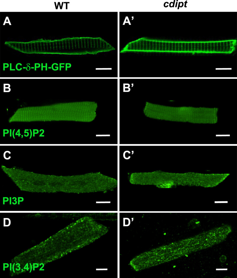
Fig 5 Localization of PIPs by immunofluorescence in wildtype and <italic>cdipt</italic> mutant zebrafish.

Confocal micrographs showing localization of PIPs is not affected in early larval development of cdipt mutants. (A, A’) visualization of skeletal muscle from live embryos injected with PLCδPH-GFP, a marker for PI(4,5)P2. There was no obvious difference in expression between wild type (WT) and cdipt mutant embryos. (B-D, B’-D’) Immunostaining with PIP antibodies of myofibers isolated from WT and cdipt mutants. Localization of PI(4,5)P2(B, B’), PI3P (C, C’) and PI(3,4)P2(D, D’) is similar in wildtype and cdipt zebrafish. Scale bars = 10μm.

Figure Data
Acknowledgments
This image is the copyrighted work of the attributed author or publisher, and ZFIN has permission only to display this image to its users. Additional permissions should be obtained from the applicable author or publisher of the image. Full text @ PLoS One