
|
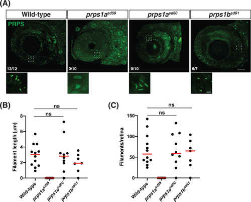
Fig. 5
Truncation of prps1a prevents phosphoribosyl pyrophosphate synthetase (PRPS) filament formation. A, Representative maximum intensity projections of confocal images from wild-type, prps1a mutant, and prps1b mutant eyes at 5 dpf, stained for PRPS. Rostral is to the left and dorsal is to the top; zoomed views of boxed areas are displayed below each panel. Fractions indicate the number of embryos exhibiting PRPS filaments out of the number of embryos examined per genotype. Only prps1asd59 mutants failed to form PRPS filaments in the eye. Scale bars: 40 μm for main images, 3 μm for zoomed images. B, Graph plots the average length of the PRPS filaments observed in each wild-type, prps1a mutant, and prps1b mutant eye at 5 dpf. Red lines indicate median values (ns, no statistically significant difference between data sets). C, Graph plots the number of PRPS filaments found in each wild-type, prps1a mutant, and prps1b mutant eye at 5 dpf. Red lines indicate median values (ns, not significant)