
Fig. 2
- ID
- ZDB-IMAGE-200814-2
- Publication
- Tao et al., 2020 - LIN28B regulates transcription and potentiates MYCN-induced neuroblastoma through binding to ZNF143 at target gene promotors
- All Figures
- Figures for Tao et al., 2020
|
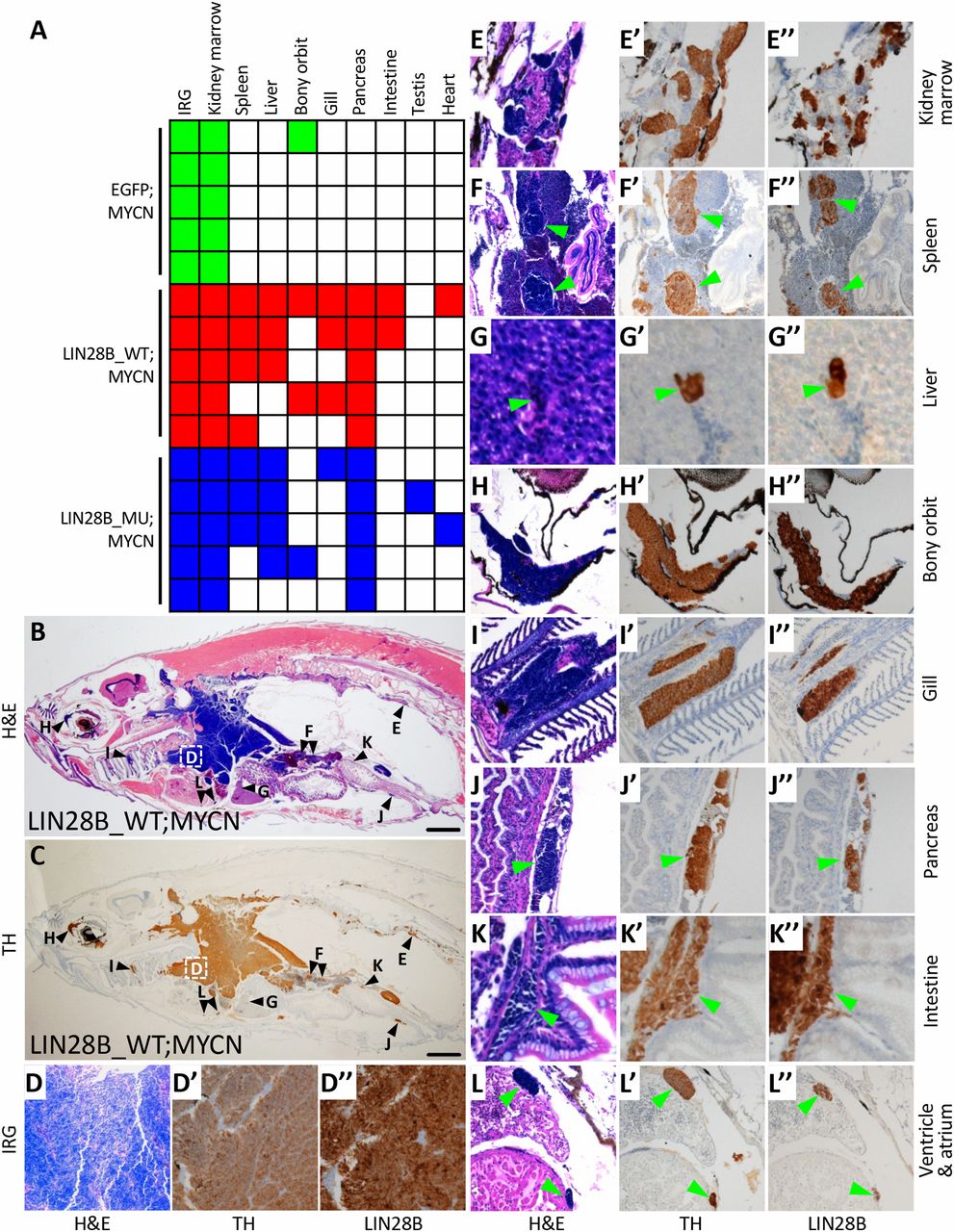
Fig. 2 LIN28B promotes the metastasis of MYCN-induced neuroblastoma. (A) Summary of neuroblastoma metastases in EGFP;MYCN, LIN28B_WT;MYCN, and LIN28B_MU;MYCN transgenic lines at 6 mo of age, five fish per group. Colored boxes indicate that metastasis was observed in the indicated organ while white boxes indicate no apparent metastasis. The difference in metastases between EGFP;MYCN and either LIN28B_WT;MYCN or LIN28B_MU;MYCN fish (P = 0.0476) was compared using the two-tailed Fisher’s exact test. (B and C) Representative images of neuroblastoma metastases in the sagittal sections of LIN28B_WT;MYCN fish by H&E staining (B) and immunohistochemical staining of TH (C). The organs harboring metastatic tumor cells are marked in B and C and are correspondingly magnified in D–L″. (Scale bar, 1 mm.) (D–L″) Magnified views of the organs shown in B and C showing H&E staining as well as immunostaining of TH and LIN28B in LIN28B_WT;MYCN fish. Green arrowheads indicate metastatic cells.