
|
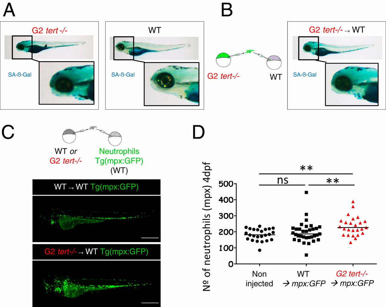
Fig. 5 G2 tert mutant cells induce high levels of senescence and systemic inflammation in host larvae. (A) Representative images of SA-β-Gal assay comparing WT and G2 tert−/− 4-dpf zebrafish embryos. Yolk sack staining is nonspecific. (B, Left) Scheme of chimera generation: G2 tert−/− blastula cells are transplanted into WT embryos (G2 tert−/− → WT). (B, Right) Marked senescence in 4-dpf WT embryos upon G2 tert−/− cell injection (C, Left) Scheme of chimera generation: WT or G2 tert−/− blastula cells transplanted into WT Tg(mpx:GFP) embryos with GFP-labeled neutrophils: WT → WT Tg(mpx:GFP) vs. G2 tert−/− → WT Tg(mpx:GFP). (C, Right) Representative images at 4 dpf, and neutrophils are shown in green. (Scale bars: 0.5 mm.) (D) Quantification of neutrophils at 4 dpf. Noninjected Tg(mpx:GFP) were used as controls. Each data point represents one zebrafish (**P > 0.01; noninjected n = 24; WT n = 33; tert−/− n = 25; ns, nonsignificant [P > 0.05]).