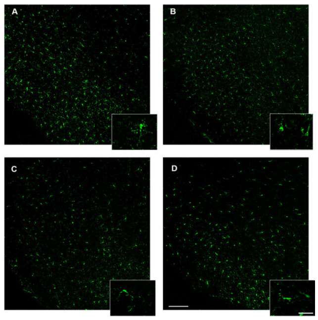
IMAGE

Figure 8

ID
ZDB-IMAGE-200811-43
Source
Figures for Imperatore et al., 2020
Image
Figure Caption

Figure 8

Iba1 immunostaining in the brain of zebrafish. (A) Representative image of the hypothalamus of control zebrafish showing resting microglia depicted in the box with high magnification. (B) Representative image of D.I.O. (diet-induced obesity zebrafish) hypothalamus showing activated or dystrophic microglia depicted in the box with high magnification. (C) Representative image of the hypothalamus of D.I.O. followed by 3,5-T2 showing activated or dystrophic microglia depicted in the box with high magnification. (D) Representative image of the hypothalamus of D.I.O. treated with 3,5-T2 showing activated or dystrophic microglia depicted in the box with high magnification. Scale bar: 100 μm in the low magnification and 25 μm in the higher magnification in the boxes.

Acknowledgments
This image is the copyrighted work of the attributed author or publisher, and ZFIN has permission only to display this image to its users. Additional permissions should be obtained from the applicable author or publisher of the image. Full text @ Animals (Basel)