
Fig. 4
|
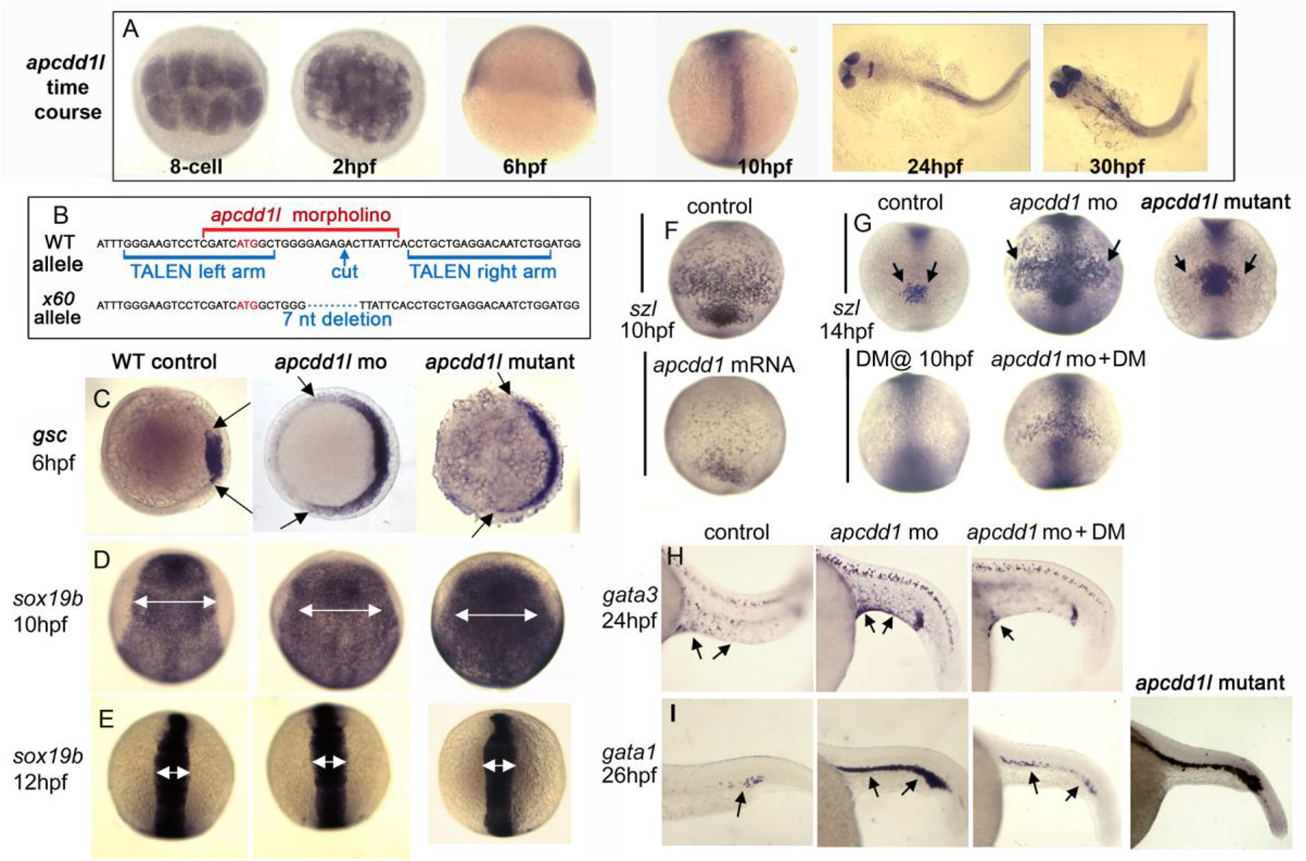
Fig. 4 Role of APCDD1 in zebrafish axis elongation and dorso-ventral patterning. A. Expression of apcdd1l in zebrafish embryos at the indicated times. The presence of maternal apcdd1l mRNA, confirmed by RT-PCR (not shown), is seen in all cells in the early blastula (animal pole views). By early gastrulation (6 hpf), apcdd1l is only weakly expressed with preferential staining in dorsal tissues (lateral view, dorsal to the right). By the end of gastrulation (10 hpf), apcdd1l is expressed throughout to the dorsal axis (dorsal view and lateral view, anterior up). At 24 and30 hpf, apcdd1l is expressed in the eyes, restricted regions in the brain, and dorsal tissues in the trunk and tail. B. Partial sequence of exon 1 surrounding the initiation codon (red font) showing the binding sites for apcdd1l-mo (red font) and TALEN left and right arms (blue font) used to induce double strand breaks. The induced x60 mutant allele has a 7 nucleotide deletion, leading to a frame shift followed by 18 premature stops in exon 1 (a presumptive null). C. Expression of the organizer gene goosecoid (gsc) at 6 hpf (early gastrula stage). Knockdown of apcdd1l, or loss of apcdd1l in MZmutants, disinhibits maternal Wnt, resulting in expansion of the organizer. Animal pole views with dorsal to the right. D-E. Expression of neural plate marker sox19 b at the end of gastrulation (C) and at the 6-somite stage (D). The early expansion of gsc in apcdd1-morphants and MZ mutants results in a dorsalized phenotype that persists through the end of gastrulation, but dorsalization is rapidly reversed by early somitogenesis stages. Dorsal views with anterior to the top. F. Expression of the Bmp feedback inhibitor sizzled (szl) in ventral ectoderm (anterior up) at the end of gastrulation. Expression of szl marks cells experiencing active Bmp signaling. Injection of apcdd1l mRNA into wild-type embryos at the one-cell stage results in downregulation of szl by the end of gastrulation. G. Expression of szl in ventral ectoderm (anterior up) at the 10-somite stage. apcdd1l-morphants and MZmutants exhibit an expanded ventral domain of szl, indicating that Bmp signaling is now higher than normal. Treatment of embryos from 10 hpf with 75 μM dorsomorphin (DM) abolishes szl expression in control embryos and partially reverses expansion of szl expression in apcdd1l-morphants. H-I. Expression of gata3 in ventral epidermis ionocytes (arrows) at 24 hpf (G) and gata1 in blood progenitors (arrows) at 27 hpf (H). Ventral ionocytes and blood progenitors are expanded in apcdd1l-morphants, indicating ventralization of caudal structures. MZapcdd1l mutants also show an expanded domain of gata1. Treatment of apcdd1l-morphants with 75 μM DM from the end of gastrulation partially reverses ventralization. Images show lateral views with anterior to the left.At least n = 15 zebrafish embryos were examined for each time point and experimental condition.
Reprinted from Developmental Biology, 464(1), Vonica, A., Bhat, N., Phan, K., Guo, J., Iancu, L., Weber, J.A., Karger, A., Cain, J.W., Wang, E.C.E., DeStefano, G.M., O'Donnell-Luria, A.H., Christiano, A.M., Riley, B., Butler, S.J., Luria, V., Apcdd1 is a dual BMP/Wnt inhibitor in the developing nervous system and skin, 71-87, Copyright (2020) with permission from Elsevier. Full text @ Dev. Biol.