
|
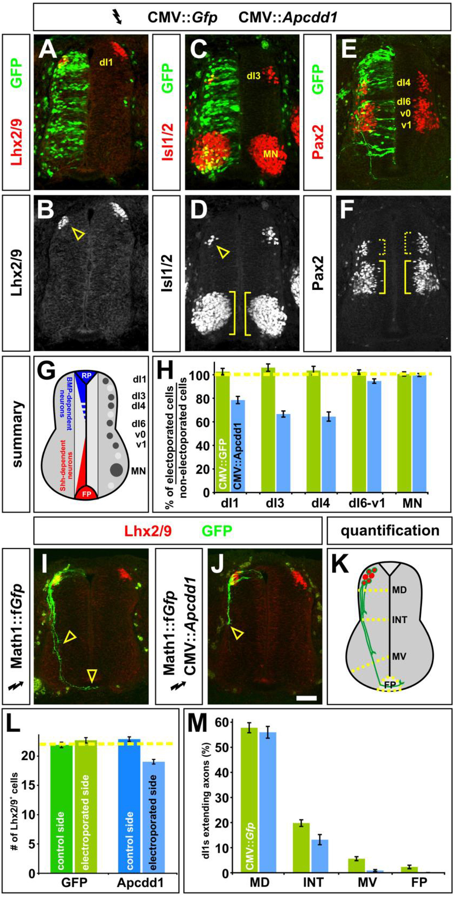
Fig. 6 Mis-expression of Apcdd1 blocks BMP-specific activities in the dorsal spinal cord. A-F, H. Gfp alone (control, H), or Gfp (green) in combination with APCDD1 (experimental, A-F, H), was ectopically expressed throughout the spinal cord under the control of the CMV enhancer by in ovo electroporation at Hamilton Hamburger (HH) stages 14/15. Embryos were harvested at HH stage 23 and examined for the number of Lhx2/9+ dI1 (commissural) neurons (red, A, B), Isl1/2+ dI3 and motor neurons (MNs) (red, C, D) or Pax2+ dI4 and dI6-v1 neurons (red, E, F). G. The relative positions of the labeled post-mitotic neurons are shown schematized within the spinal cord. The dorsal most populations of spinal neurons (dI1-dI3) are known to be dependent on BMP signaling, the role of BMP signaling in the more ventral-dorsal populations (dI4-dI6) has remained unresolved. H. Quantification revealed that the misexpression of Gfp (n = 42 sections from 6 embryos) had no significant effect on any of the monitored populations of spinal neurons (dI1, p > 0.64 compared to the non-electroporated control side of the same embryo; dI3, p > 0.29; dI4, p > 0.44; dI6-v1, p > 0.40; MN, p > 0.49). In contrast, the ectopic expression of both Gfp and Apcdd1 (n = 35 sections from 6 embryos) resulted in a 25–35% loss in BMP-dependent dorsal neural populations: dI1 neurons (p < 0.03) dI3 neurons (p < 0.008) and dI4 neurons (p < 3.5 × 10⁻⁶). However, APCDD1 had no detectable effect on the numbers of BMP-independent neurons, specifically dI6, vo, v1 interneurons (p > 0.055) and MNs (p > 0.54). I. Lhx2/9+ dI1 neurons (red) electroporated with farnesylated (f) Gfp under the control of the Math1 (Atoh1) enhancer (Math1::fGfp) at HH stages 16/17, GFP+ axons (green) are extending into the ventral spinal cord by HH stage 22, with a few axons having reached the floor plate (FP, arrowheads). J. In contrast, Lhx2/9+ dI1 neurons (red) concomitantly electroporated with both Math1::fGfp (green) and CMV:: Apcdd1 showed over an 80% reduction of axon outgrowth at the same stage, with no axons reaching the FP (arrowhead). K. The extent of the dI1 axon outgrowth was quantified by determining whether dI1 axons had crossed one of four arbitrary lines in the spinal cord: mid-dorsal (MD), intermediate (INT), mid-ventral (MV) or the FP, as in (Phan et al., 2010). L. Misexpression of Apcdd1 throughout the spinal cord at HH stage 16/17 has a small but significant effect on the numbers of dI1 neurons. There is a 10% reduction in dI1 neurons on the APCDD1-electroporated side (p < 2.2 × 10⁻⁹, n = 173 sections from 6 embryos) compared to the control Gfp electroporated side (n = 178 sections from 5 embryos). M. By HH stage 22, approximately 55% of Lhx2/9+ neurons electroporated with gfp (control) or gfp with APCDD1 (experimental) have extended GFP+ axons. In control embryos (n = 178 sections, taken from 5 embryos), 10% of these axons extended to the MV line and 4% reached the FP. In contrast, only 1.6% of Lhx2/9+ neurons in the embryos electroporated with both Gfp and Apcdd1 (n = 173 sections, taken from 6 embryos) had extended to axons to the MV line (p < 0.0001, similar to GFP controls) with none reaching the FP (p < 0.02). Statistical analyses were performed using a one-tailed Student’s t-test, when the data fit a normal distribution, and with the Mann–Whitney test. Scale bar: 25 μm.
Reprinted from Developmental Biology, 464(1), Vonica, A., Bhat, N., Phan, K., Guo, J., Iancu, L., Weber, J.A., Karger, A., Cain, J.W., Wang, E.C.E., DeStefano, G.M., O'Donnell-Luria, A.H., Christiano, A.M., Riley, B., Butler, S.J., Luria, V., Apcdd1 is a dual BMP/Wnt inhibitor in the developing nervous system and skin, 71-87, Copyright (2020) with permission from Elsevier. Full text @ Dev. Biol.