
Image
|
Figure Caption
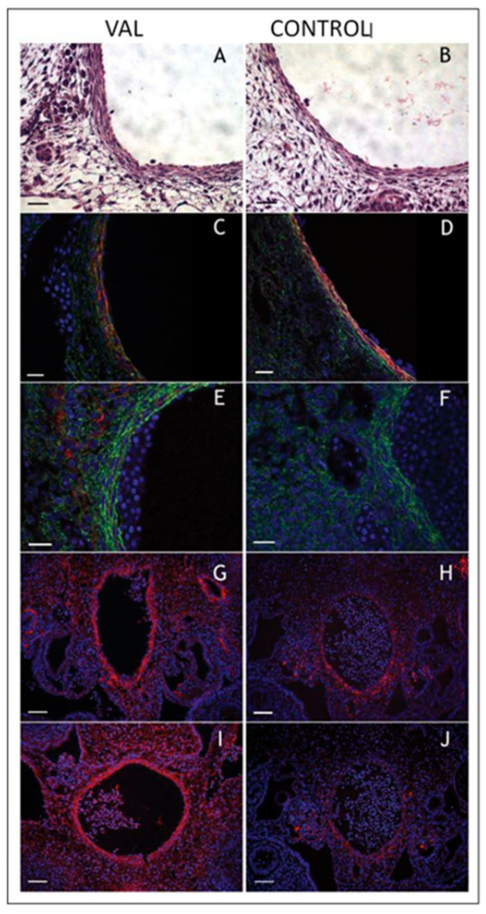
Figure 14
Increased arterial load via unilateral vitelline artery ligation (VAL) alters aortic structural properties. Representative images of VAL (
Acknowledgments
This image is the copyrighted work of the attributed author or publisher, and
ZFIN has permission only to display this image to its users.
Additional permissions should be obtained from the applicable author or publisher of the image.
Full text @ J Cardiovasc Dev Dis