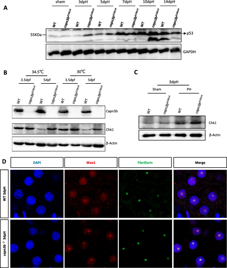
IMAGE

Fig. 9

ID
ZDB-IMAGE-200629-25
Genes
Antibodies
Source
Figures for Chen et al., 2020
Image
Figure Caption

Fig. 9

Hepatic accumulation of Chk1 and Wee1 in capn3bΔ19Δ14 at 3dpH. a Elevation of p53 protein level in capn3bΔ19Δ14 after PH. Western blot analysis of p53 at different time point after PH as indicated. Total proteins were extracted from the liver tissues of sham, 3dpH, 5dpH, 7dpH, 10dpH and 14dpH, respectively. GAPDH, loading control. b Western blot of Capn3b and Chk1 in WT and capn3bΔ19Δ14 mutant embryos at 3.5dpf and 5dpf grown at 30 °C and 34.5 °C, respectively. The embryos were shifted to 34.5 °C at 12hpf till the time of harvesting. β-Actin: loading control. c Western blot of Chk1 in WT and capn3bΔ19Δ14 sham and PH groups at 3dpH group. Fibrillarin: loading control. d Co-immunostaining of Wee1 and Fibrillarin in the liver of WT and capn3bΔ19Δ14 mutant fish at 3dpH. DAPI: staining nuclei

Figure Data
Acknowledgments
This image is the copyrighted work of the attributed author or publisher, and ZFIN has permission only to display this image to its users. Additional permissions should be obtained from the applicable author or publisher of the image. Full text @ Cell Regen (Lond)