
Fig. 8
|
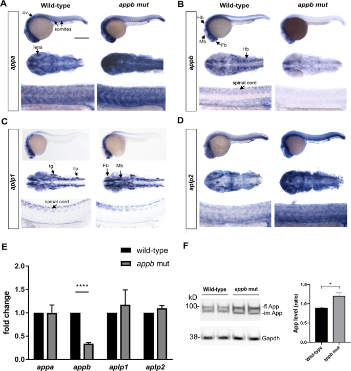
Fig. 8
Expression of app family in appb mutants. Whole mount in situ hybridization of appa (A), appb (B), aplp1 (C) and aplp2 (D) at 24 hpf. Upper panel: whole embryos; middle panel: head (dorsal view) and lower panel: trunk in each set of genes (anterior to left). Scale bar =500 μm (upper panel), 100 μm (middle panel) and 50 μm (lower panel). (E) Whole embryo qPCR analysis of appa, appb, aplp1 and aplp2 at 24 hpf. Values are reported as mean ±SEM. ****p < 0.001. (F) Western blot analysis and quantification of zebrafish App expression in appb mutants (n = 4) at 24 hpf using the Y188 antibody shown as ratio of wild-type (n = 4). Mean is reported as ± SD. Fl, full length; im, immature. *p < 0.05, **p < 0.01 and ****p < 0.001. ov, otic vesicle; Fb, forebrain; Mb, midbrain; Hb, hindbrain; llp, lateral line primordium; tg, trigeminal ganglia.