
|
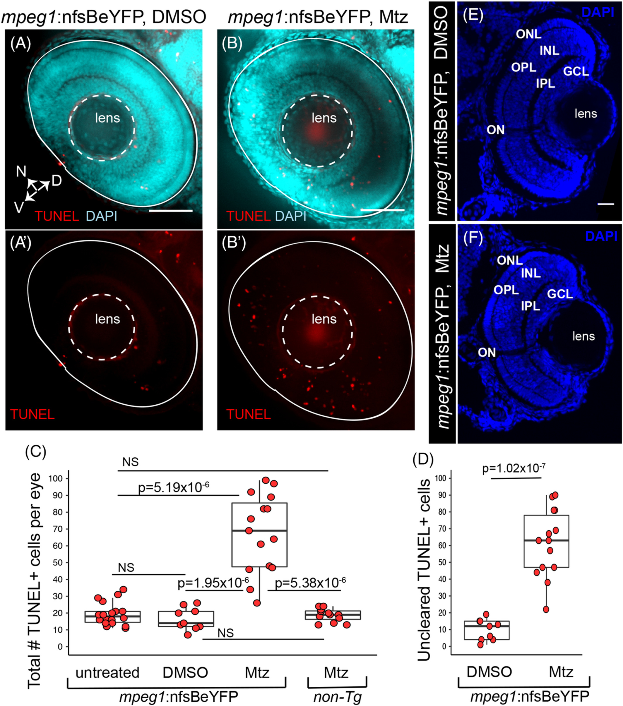
Fig. 2 Accumulation of TUNEL+ cells in the retina when primitive microglia are depleted. Whole embryos were collected and processed for TUNEL (red) staining then DAPI (cyan) counterstaining. Images show selected z projections of whole eyes from mpeg1 :nfsBeYFP embryos treated with DMSO, (A, A′), or Mtz, (B, B′). Orientation is indicated by the compass on bottom left of panel A, N = nasal, V = ventral, D = dorsal (applies to A‐B′). The lens and boundary of the eye are indicated by white dashed outlines. C, The total number of TUNEL+ cells per eye was quantified for each indicated group. Box plots are shown for each group with each individual data point shown in red circles. A Welch's one‐way test (P = 7.27 × 10⁻⁷) was performed, followed by Bonferroni post hoc test. P values shown for pairwise comparison with P < .05; NS = not significant. D, Since TUNEL signal is often associated with YFP+ microglia (see Figure 1A′,B′), we determined the number of uncleared TUNEL+ cells in mpeg1 :nfsBeYFP retinas treated with DMSO or Mtz by subtracting the number of microglia‐associated TUNEL+ puncta from the total number of TUNEL+ cells. Box plots are shown for each group with each individual data point shown in red circles. P ‐value (Welch's t test) is shown. E and F, Comparison of gross retinal structure from retinal cryosections from control (D) or microglia depleted (E) eyes. DAPI (blue) was used to label cell nuclei and assess retinal structure. Stereotypical retinal structure is apparent for both control (E) and microglia‐depleted (F) retinas as evidenced by organized outer nuclear layer (ONL), inner nuclear layer (INL), and ganglion cell layer (GCL). The outer and inner plexiform layers (OPL and IPL, respectively) and location of the optic nerve (ON) are also apparent. Scale bar in E = 20 μm, applies to E and F. Samples sizes: mpeg1 :nfsBeYFP untreated = 18 eyes from 15 fish, mpeg1 :nfsBeYFP DMSO treated = 9 eyes from 5 fish, mpeg1 :nfsBeYFP Mtz treated = 14 eyes from 10 fish, Non‐Tg Mtz treated = 12 eyes from 6 fish