IMAGE

Figure 1

ID
ZDB-IMAGE-200608-11
Genes
Source
Figures for Li et al., 2020
Image
Figure Caption

Figure 1

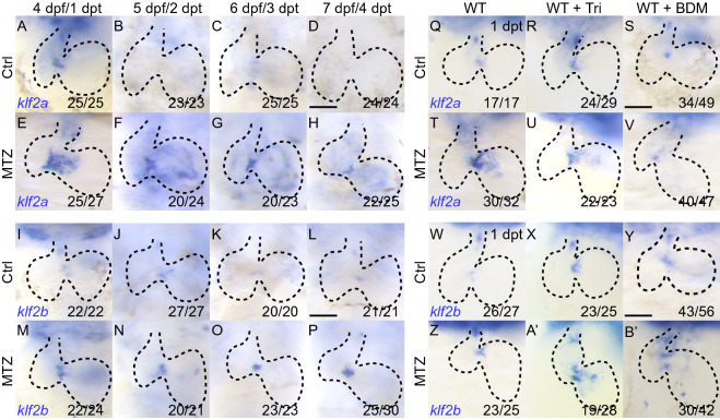
Reduced blood flow changesklf2aandklf2bexpression during ventricle regeneration. (A–H) Whole-mount in situ hybridizations showing klf2a upregulation in ablated hearts (E–H) compared to control hearts (A–D) at 4–7 dpf/1–4 dpt. (I–P) Whole-mount in situ hybridizations showing klf2b upregulation in ablated hearts (M-P) compared to control hearts (I–L) at 4–7 dpf/1–4 dpt. (Q–V) Whole-mount in situ hybridizations indicated klf2a upregulation after ablation (Q, T) was blocked in Tricaine treated hearts (R, U) and BDM treated hearts (S, V) at 1 dpt. (W–B’) Whole-mount in situ hybridizations indicated klf2b upregulation after ablation (W, Z) was slightly enhanced in Tricaine treated hearts (X, A’) and BDM treated hearts (Y, B’) at 1 dpt. Scale bars, 50 µm. dpt, days post treatment. Dashed lines outline the hearts. Numbers indicate the ratio of representative staining observed

Figure Data
Acknowledgments
This image is the copyrighted work of the attributed author or publisher, and ZFIN has permission only to display this image to its users. Additional permissions should be obtained from the applicable author or publisher of the image. Full text @ Protein Cell