IMAGE

Figure 6.

ID
ZDB-IMAGE-200605-7
Source
Figures for He et al., 2020
Image
Figure Caption

Figure 6.

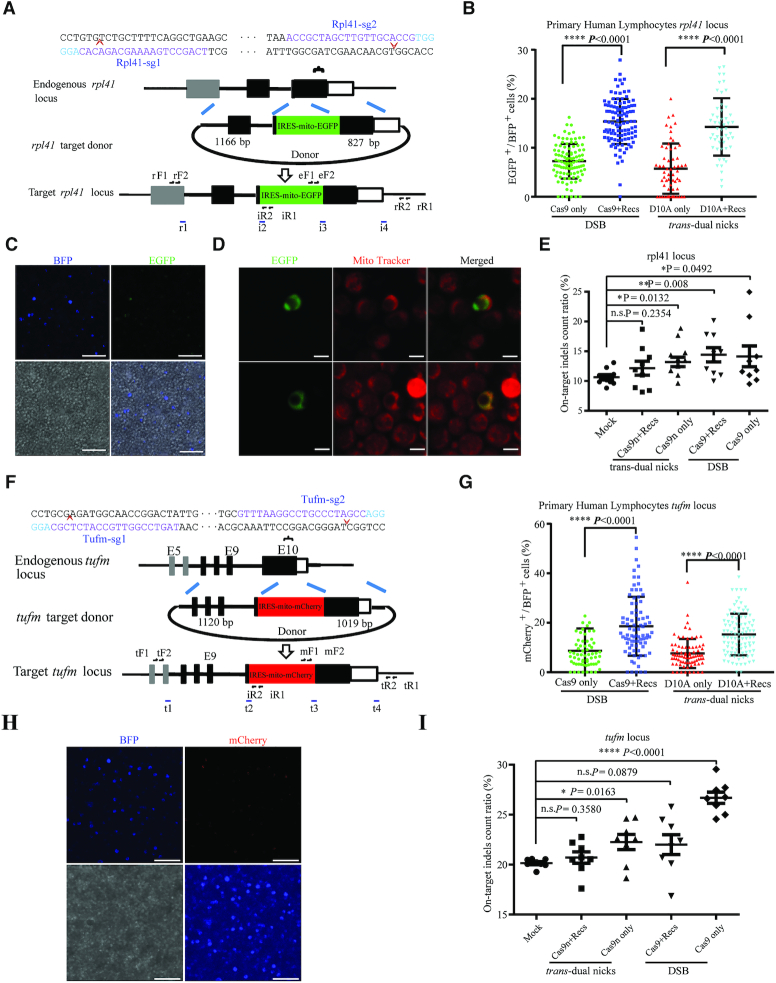
NEO achieved efficient integration in non-dividing primary human PBLCs. (A) Schematic overview of the strategy to generate the Rpl41-IRES-mito-EGFP cells. rF1, rF2, iR1, rR2, i2, i3, i4, eF1 and eF2 are the nest PCR primers (Supplementary Table S4). (B) Relative integration efficiencies (EGFP+ / BFP+) with IRES-mito-EGFP precise integration at the Rpl41 locus. Results were obtained from at least 2000 BFP+ cells and three independent electroporation experiments. The input data points calculated from each randomly selected image field were shown as dots. ****P < 0.0001, Student's t-test. (C) Mito-EGFP signals in the Rpl41-KI positive hPBLCs. Cells in blue were positively transfected with TagBFP that served as an indicator. Scale bar, 50 μm. (D) Live staining of hPBLCs with mito Tracker Red. Green, Rpl41-KI positive hPBLCs. MitoTracker Red fluorescence dye specifically labelled mitochondria in red. Scale bar, 5 μm. (E) On-target indel count ratio detected by IDAA at the Rpl41 locus. Total indel count ratio was calculated as the summed peak areas of all indel peaks relative to the total peak area. Results were obtained from at least three samples from independent electroporation experiments, each sample was tested three times. Mock, control group with mock treatment. n.s., no significant difference. *P < 0.05, **P < 0.01, Student's t-test. Error bars are s.d. (F) Schematic overview of the strategy to generate a TUFm-IRES-mito-mCherry KI cell. The nested PCR primers used for knock-in identification are shown (Supplementary Table S4). (G) Relative efficiencies of IRES-mito-mCherry precise integration at the TUFm locus. Results were obtained from at least 2000 BFP+ cells and three independent electroporation experiments. The input data points calculated from each randomly selected image field were shown as dots. ****P < 0.0001, Student's t-test. (H) Mito-mCherry signals in TUFm-KI positive hPBLCs. Cells in blue were positively transfected with TagBFP mRNA. Scale bar, 50 μm. (I) On-target indels count ratio detected by IDAA at the TUFm locus. Results were obtained from at least three samples from independent electroporation experiment, each sample was tested three times. Mock, control group with mock treatment. n.s., no significant difference, *P < 0.05, ****P < 0.0001, Student's t-test. Error bars denote s.d.

Acknowledgments
This image is the copyrighted work of the attributed author or publisher, and ZFIN has permission only to display this image to its users. Additional permissions should be obtained from the applicable author or publisher of the image. Full text @ Nucleic Acids Res.