
Fig. 5
- ID
- ZDB-IMAGE-200605-25
- Publication
- Xia et al., 2020 - ube3d, a New Gene Associated with Age-Related Macular Degeneration, Induces Functional Changes in Both In Vivo and In Vitro Studies
- All Figures
- Figures for Xia et al., 2020
|
Fig. 5
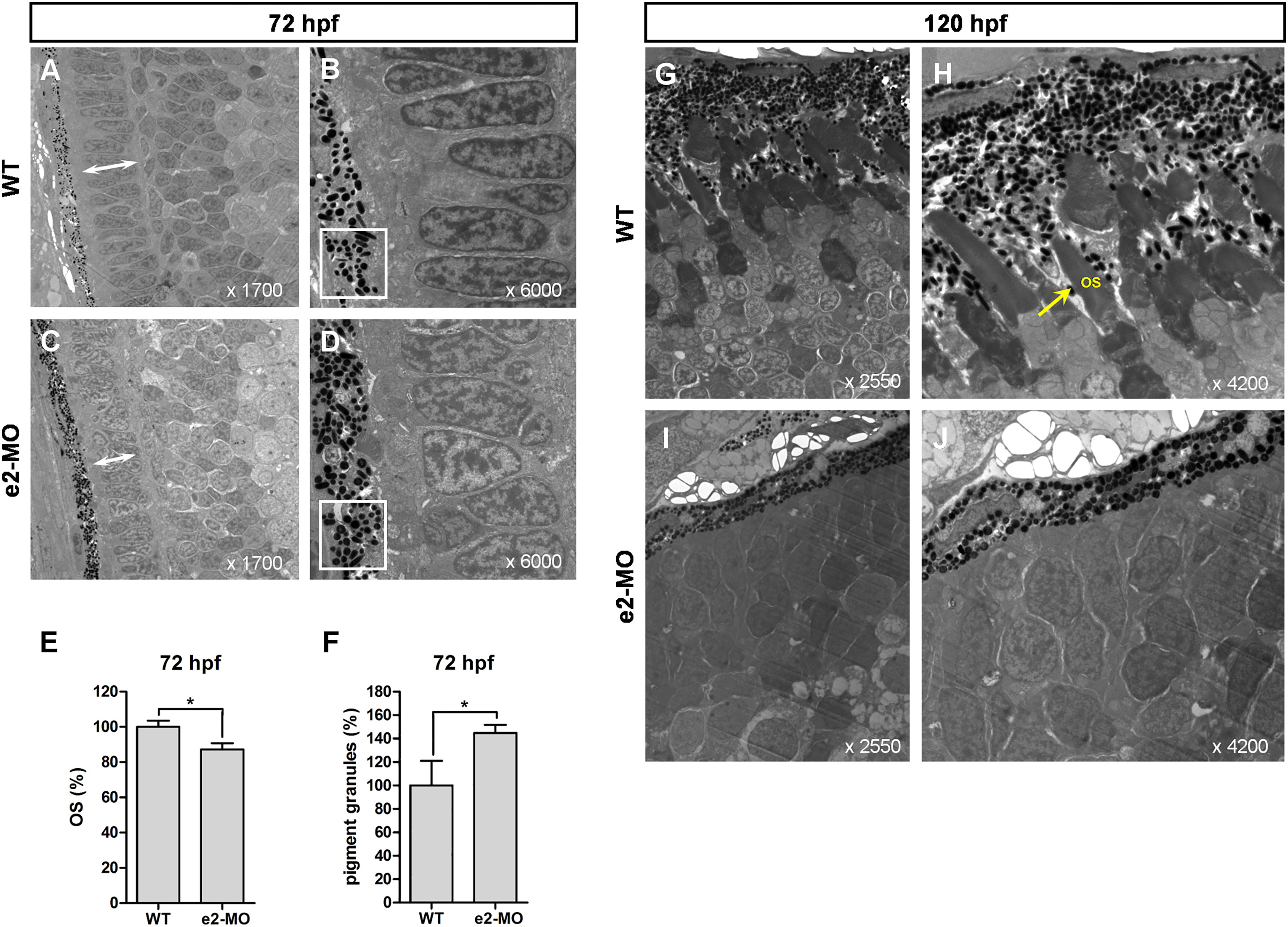
Knockdown of ube3d Results in More Deposited Pigment Granules and an Abnormal Photoreceptor-Outer Segment Layer
(A) Transmission electron micrographs of transverse sections cut along the dorsal-ventral axis of WT zebrafish eyes at 72 hpf (with ×1,700 and ×6,000 magnification, as indicated). (B) Enlargement of (A). (C) Transmission electron micrographs of transverse sections cut along the dorsal-ventral axis of e2-MO ube3d morphant eyes at 72 hpf (with ×1,700 and ×6,000 magnification, as indicated). (D) Enlargement of (C). (E) Graphical representation demonstrating that OS (arrow) lengths were shorter in the ube3dmorphants than those in the WT larvae. (F) Graphical representation demonstrating that more pigment granules were deposited in the photoreceptor OS layer in ube3dmorphants than were deposited in WT larvae. (G) Photoreceptor OSs (arrow) were present in WT 120-hpf larvae (with ×2,550 and ×4,200 magnification, as indicated). (H) Enlargement of (G). (I) Photoreceptor OSs were not observed in the ube3d morphants at 120 hpf (with ×1,700 and ×6,000 magnification, as indicated). (J) Enlargement of (I). The data are presented as the mean ± SD. ∗p < 0.05.