
|
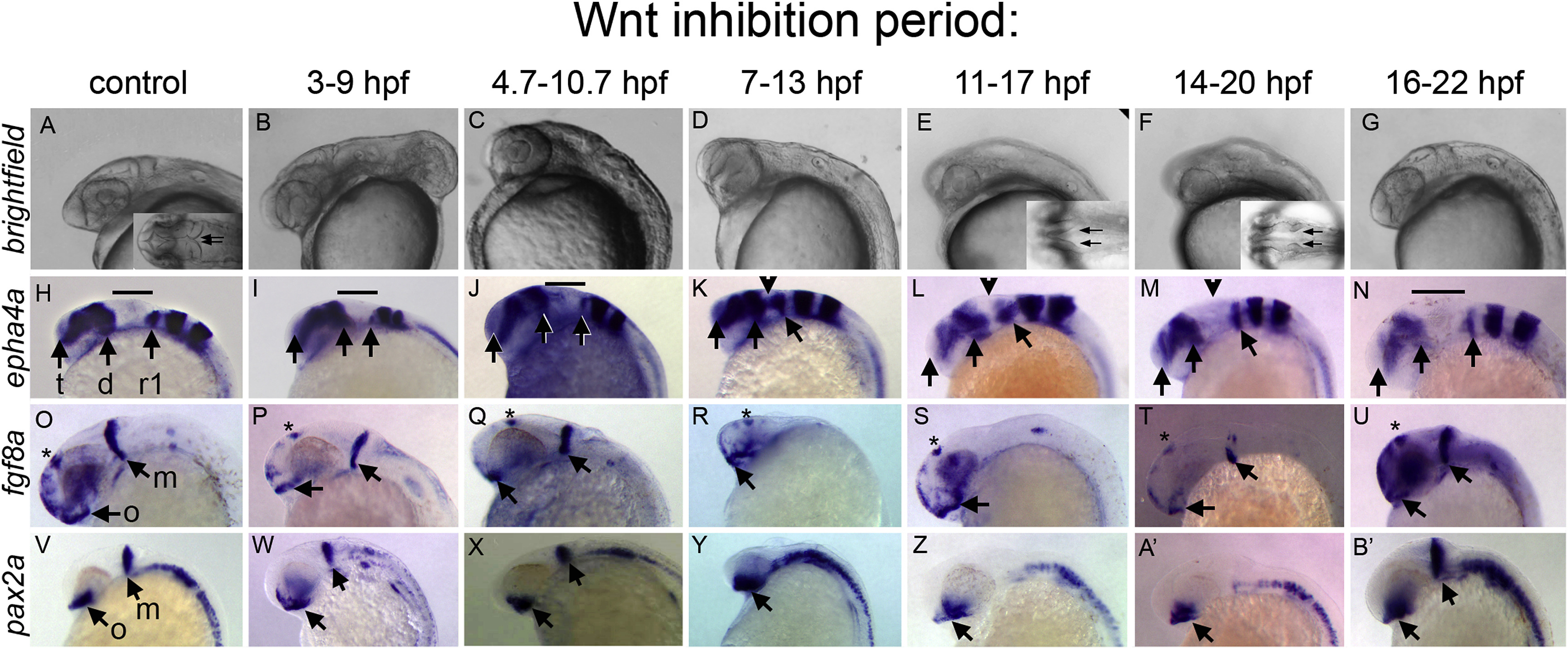
Fig. 1 Timed inhibition of Wnt/β-catenin signaling reveals distinct neural plate responses. Embryos derived from hs:dkk1b/+ outcrosses were heat shocked to inhibit Wnt signaling during the time periods indicated above each column and in Fig. S2. All images: heads of 27 hpf embryos, anterior left, dorsal up. Brightfield (A–G). Insets in (A,E,F) are dorsal views, arrows indicate inner edge of MHB constrictions, which touch in control embryos. In situ hybridizations to epha4a (H–N), fgf8a (O–U), and pax2a (V–B’). Wild-type siblings to hs:dkk1b/+ embryos are shown in control (A,H,O,V). Note that Wnt inhibition between 3-9 hpf produces a range of dorsalized phenotypes; C3 dorsalized embryos are shown in (B,I,P,W). In situ results are equivalent in all dorsalized classes. (H–N) Arrows indicate telencephalon (t), diencephalon (d), rhombomere 1 (r1). Bar indicates midbrain, MHB and cerebellar primordia that do not express epha4a, indicated with arrowheads in (K,L,M). (O–B’) Arrows indicate optic stalk (o), and mhb (m, when present). Asterisks in (O–U) indicate dorsal diencephalon domain.
Reprinted from Developmental Biology, 462(2), Green, D.G., Whitener, A.E., Mohanty, S., Mistretta, B., Gunaratne, P., Yeh, A.T., Lekven, A.C., Wnt signaling regulates neural plate patterning in distinct temporal phases with dynamic transcriptional outputs, 152-164, Copyright (2020) with permission from Elsevier. Full text @ Dev. Biol.